This site uses cookies to improve your experience. To help us insure we adhere to various privacy regulations, please select your country/region of residence. If you do not select a country, we will assume you are from the United States. Select your Cookie Settings or view our Privacy Policy and Terms of Use.
Cookie Settings
Cookies and similar technologies are used on this website for proper function of the website, for tracking performance analytics and for marketing purposes. We and some of our third-party providers may use cookie data for various purposes. Please review the cookie settings below and choose your preference.
Used for the proper function of the website
Used for monitoring website traffic and interactions
Cookie Settings
Cookies and similar technologies are used on this website for proper function of the website, for tracking performance analytics and for marketing purposes. We and some of our third-party providers may use cookie data for various purposes. Please review the cookie settings below and choose your preference.
Strictly Necessary: Used for the proper function of the website
Performance/Analytics: Used for monitoring website traffic and interactions
This was sent by anonymous The patient is a 55-year-old male who presented to the emergency department after approximately 3 to 4 days of intermittent central boring chestpain initially responsive to nitroglycerin, but is now more constant and not responsive to nitroglycerin. It is unknown when this pain recurred and became constant.
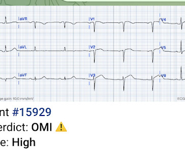
Written by Jesse McLaren A healthy 75 year old developed 7/10 chestpain associated with diaphoresis and nausea, which began on exertion but persisted. Below is the first ECG recorded by paramedics after 2 hours of chestpain, interpreted by the machine as “possible inferior ischemia”. What do you think?
A 56 year old male with PMHx significant for hypertension had chestpain for several hours, then presented to the ED in the middle of the night. He reported chestpain that developed several hours prior to arrival and was 5/10 in intensity. The pain was located in the mid to left chest and developed after riding his bike.
Written by Colin Jenkins and Nhu-Nguyen Le with edits by Willy Frick and by Smith A 46-year-old male presented to the emergency department with 2 days of heavy substernal chestpain and nausea. The patient continued having chestpain. These diagnoses were not found in his medical records nor even a baseline ECG.
Written by Willy Frick A man in his 50s with history of hypertension, hyperlipidemia, and a 30 pack-year smoking history presented to the ER with 1 hour of acute onset, severe chestpain and diaphoresis. preceding each of the fascicular beats — indicating a faster rate for the escape rhythm compared to the sinus bradycardia ).
No ChestPain, but somnolent. The fact that this is syncope makes give it a far lower pretest probability than chestpain, but it was really more than syncope, as the patient actually underwent CPR and had hypotension on arrival of EMS. Here is the ED ECG (a photo of the paper printout) What do you think?
See our other countless hyperkalemia cases below: General hyperkalemia cases: A 50s year old man with lightheadedness and bradycardia Patient with Dyspnea. A woman with near-syncope, bradycardia, and hypotension What happens if you do not recognize this ECG instantly? Also: How did this happen? Is this just right bundle branch block?
A 50-something man presented in shock with severe chestpain. Here is his ED ECG: There is bradycardia with a junctional escape. Case continued A bedside ultrasound showed diminished LV EF and of course bradycardia. His prehospital ECG was diagnostic of inferior posterior OMI. He appeared gray in color, with cool skin.
Case An 82 year old man with a history of hypertension presented to the ED with chestpain at 1211. He described his chestpain as pleuritic and reported that it started the day prior while swinging a golf club. His pain suddenly became much worse in the ED and he became acutely diaphoretic, dizzy, and hypotensive.
This was sent to me by a former resident from a community hospital: A middle-aged woman complained of chestpain and was seen in triage. The computer interpreted the ECG (GE Marquette 12 SL) as: "Sinus Bradycardia. Here it is: Computer interpretation: "Sinus bradycardia. She had a ECG recorded. Normal ECG."
The patient presented due to chestpain that was typical in nature, retrosternal and radiating to the left arm and neck. He denied any exertional chestpain. It is unclear if the patient was pain free at this time. He has a medical hx notable for hypertension, hyperlipidemia and previous tobacco use disorder.
The patient presented with chestpain. I was taught that the tell-tale sign of ischemia vs an electrical abnormality was in the hx, i.e. chestpain for the ischemia and potential syncope for brugada. Only 5-18% of ED patients with chestpain have a myocardial infarction of any kind. Bradycardia.
Submitted and written by Megan Lieb, DO with edits by Bracey, Smith, Meyers, and Grauer A 50-ish year old man with ICD presented to the emergency department with substernal chestpain for 3 hours prior to arrival. At this time he reported ongoing chestpain and was given aspirin and nitroglycerin.
Written by Jesse McLaren, with edits from Smith and Grauer A 60 year old with no past medical history presented with two hours of chestpain radiating to the left arm, with normal vitals. Unfortunately, the reality is — that many ( most ) WPW patients who present with chestpain do not manifest intermittent preexcitation.
The patient’s chestpain spontaneously resolved before he was evaluated and has a repeat ECG obtained at 22:12 obtained shown below. Soon afterward, the patient’s symptoms return along with lightheadedness, bradycardia, and hypotension. It is unclear if he received aspirin at triage. This ECG is more difficult.
The patient has acute chestpain. Instead — my thoughts were as follows: The rhythm is sinus , with marked bradycardia and a component of sinus arrhythmia. This was texted to me in real time. What do you think? Here was my answer: "Not ischemia. Maybe HOCM or another form of LVH. I would not activate cath lab.
I see the following: The rhythm is sinus bradycardia at ~55-60/minute. PEARL # 2: Applying PEARL #1 to today's case — the fact that this patient's symptoms began before ECG #1 was obtained, and that his chestpain had resolved by the time ECG #1 was recorded — strongly suggests that the "culprit" artery may have spontaneously opened.
The patient with no prior cardiac history presented in the middle of the night with acute chestpain, and had this ECG recorded during active pain: I did not see any ischemia on this electrocardiogram. Their apparently excessive length (QT interval) is due to bradycardia. This is a case I had quite a while back.
Written by Pendell Meyers A woman in her 50s presented with acute chestpain and lightheadedness since the past several hours. Here is her triage ECG during active symptoms: What do you think? The ED physician read this as "Normal sinus rhythm. Marked ST abnormality, possible anterior subendocardial injury."
A middle-aged woman with a history of hypertension presented with typical chestpain. Here was her presenting ECG, with chestpain: Inferior leads show hyperacute T-waves and reciprocal STD in aVL, with a reciprocally hyperacute T-wave in aVL. Her BP was 160/80. This is all but diagnostic of inferior OMI.
She was hemodynamically stable — and did not have chestpain, lightheadedness or syncope. Even if we stopped here — We could conclude the following: There is marked bradycardia in today's rhythm ( ie, Heart rate in the low 30s ). QUESTIONS: HOW would you interpret the rhythm in Figure-1 ? Is this " high -grade" AV block?
This ECG could easily be seen in an ED chestpain patient, and I have seen many) What do you think? Description Sinus bradycardia. Comment I (Smith) have seen many similar ECGs in ED chestpain patients. I have seen this innumerable times in chestpain patients in the Emergency Department.
The rhythm is sinus bradycardia at a rate just over 50/minute. Although difficult to measure ( because of marked overlap of the QRS in multiple chest leads ) — there appears to be greatly increased QRS amplitude, consistent with voltage for LVH. To improve visualization — I've digitized the original ECG using PMcardio ).
to 1828 msec. ) — which corresponds to a variation in the rate of sinus bradycardia from 36-to-33/minute. This makes sense given that the underlying rhythm in today's case appears to be marked sinus bradycardia and arrhythmia , with a ventricular escape rhythm appearing when the SA node rate drops below 33/minute.
He did not have chestpain. Chestpain in high risk patient. Syncope and Bradycardia Syncope in a 20-something woman Long QT: Do not trust the computerized QT interval when the QT is long An Alcoholic Patient with Syncope Cardiac Arrest. Here is his triage ECG: What do you think? Is it STEMI? What is going on here?
He arrived to the ED by helicopter at 1507, about three hours after the start of his chestpain while chopping wood around noon. He arrived to the ED by ambulance at 1529, only a half hour after the start of his chestpain around 1500 while eating. Angiography revealed a 30% nonobstructive stenosis of the mid LAD.
This middle aged male with h/o GERD but also h/o stents presented to the ED with chestpain. The computer called "Sinus Bradycardia" only (implying that everything else is normal. The overreading Cardiologist called it only "Sinus Bradycardia" with no other findings. There is zero ST Elevation.
The patient in today’s case is a previously healthy 40-something male who contacted EMS due to acute onset crushing chestpain. The pain was 10/10 in intensity radiating bilaterally to the shoulders and also to the left arm and neck. Written By Magnus Nossen — with edits by Ken Grauer and Smith. The below ECG was recorded.
The chestpain quickly subsided. During the night, while on telemetry, the patient became bradycardic, with periods of isorhythmic AV dissociation (nodal escape rhythm alternating with sinus bradycardia), and there were sporadic PVCs. Are you worried about OMI in this case? His response was: "I would not call it OMI.
He woke up alert and with chestpain which he also had experienced intermittently over the previous few days. The history in today's case with sudden loss of consciousness followed by chestpain is very suggestive of ACS and type I ischemia as the cause of the ECG changes. What do you think?
Later, I found old ECGs: 5 month prior in clinic: V5 and V6 look like OMI 9 months prior in clinic with no chest symptoms: V5 and V6 look like OMI 1 year prior in the ED with chestpain: V5 and V6 sure look like a STEMI For this ECG and chestpain in the ED, the Cath lab activated. But the angiogram was clean.
ECG of pneumopericardium and probable myocardial contusion shows typical pericarditis Male in 30's, 2 days after Motor Vehicle Collsion, complains of ChestPain and Dyspnea Head On Motor Vehicle Collision. Gunshot wound to the chest with ST Elevation Would your radiologist make this diagnosis, or should you record an ECG in trauma?
A 40-something male with no previous cardiac disease presented with chestpain. The pain continued and the first high sensitivity troponin I returned at 105 ng/L Another ECG was recorded: The ST segment in aVF has flattened a bit, revealing that there is some STD in addition to the non-specific findings in III and aVL.
All of the patients presented with chestpain , and they are all in triage. Triage is backed up, and 10 minutes into your shift one of the ED nurses brings your several ECG s that has not been overread by a physician. Which, if any, of these patients has OMI, with myocardium at risk and need for emergent PCI?
This is the initial ED ECG of a 46 year old male with chestpain: The QTc was 420 ST Elevation at 60 ms after the J-point in lead V3 = 2.5 ng/ml) A 45 year old male called 911 for chestpain: The QTc was 400 ST Elevation at 60 ms after the J-point in lead V3 = 3.5 100% LAD occlusion. He underwent CABG. QRS V2 = 15.5
He denied any chestpain or shortness of breath and stated he felt at his baseline yesterday prior to drug use. They recommended repeating his ECG and awaiting troponin since the patient did not have any chestpain. He complained of generalized weakness and left lower extremity numbness. What is it? Activate the Cath Lab?
Apparently he denied chestpain. JAMA 2000) showed that 1/3 of patients with STEMI, and 1/3 of patients with NSTEMI, present without chestpain. Chestpain and Concordant ST Depression in a patient with aortic valve and previously normal angiogram Right Bundle Branch Block and ST Depression in V1-V3.
A 55 year old with no previous cardiac history presented with 3 hours of chestpain. The pain was persistent and reportedly still present at the time of the ECG. He has now implemented the Queen of Hearts in his hospital. He wanted to share one of the first cases. This will be corrected in Version 2, coming soon.
The relationship between low RHR and CI has yet to be described.Purpose:We hypothesize that resting sinus bradycardia (low RHR) could be a predictor of chronotropic incompetence and reduced exercise capacity.Methods:The derivation cohort consists of 201 patients with normal Bruce protocol treadmill stress echocardiogram. x age in years).Results:The
If you experience any symptoms, such as chestpain, dizziness, unusual tiredness or fatigue, shortness of breath, or irregular heartbeat, your doctor would want you to go for an ECG test to find out the underlying cause. A fast heartbeat is called tachycardia, while a slow heartbeat is called bradycardia in medical terms.
That said — obvious findings include: i ) Marked bradycardia! — MY " Q uick T houghts" on Today's CASE: The ECG in Figure-1 is highly concerning — so it is indeed fortunate that this elderly woman came to the ED when she did! The rhythm in Figure-1 is complex — and defies precise interpretation without careful study. be regular! —
Written by Willy Frick A 46 year old man with a history of type 2 diabetes mellitus presented to urgent care with complaint of "chest burning." The ECG shows sinus bradycardia but is otherwise normal. The patient said his chestpain was 4/10, down from 8/10 on presentation. The following ECG was obtained.
Here are inferior leads, and aVL, magnified: A closer inspection of the inferior leads and aVL Sinus bradycardia. She went on to describe her chestpain as a "buffalo sitting on my chest" and a "weird" sensation in her jaw for 1 hour prior to arrival, associated with lightheadedness and diaphoresis. What do you think?
The combination of absence of chestpain and history of LV aneurysm made it easy to assess that this patient does not have acute OMI. In view of the History with the current admission ( ie, presenting to the ED for vertigo — with no new chestpain ) — I interpreted ECG #1 as no OMI.
We organize all of the trending information in your field so you don't have to. Join thousands of users and stay up to date on the latest articles your peers are reading.
You know about us, now we want to get to know you!
Let's personalize your content
Let's get even more personalized
We recognize your account from another site in our network, please click 'Send Email' below to continue with verifying your account and setting a password.


Let's personalize your content