This site uses cookies to improve your experience. To help us insure we adhere to various privacy regulations, please select your country/region of residence. If you do not select a country, we will assume you are from the United States. Select your Cookie Settings or view our Privacy Policy and Terms of Use.
Cookie Settings
Cookies and similar technologies are used on this website for proper function of the website, for tracking performance analytics and for marketing purposes. We and some of our third-party providers may use cookie data for various purposes. Please review the cookie settings below and choose your preference.
Used for the proper function of the website
Used for monitoring website traffic and interactions
Cookie Settings
Cookies and similar technologies are used on this website for proper function of the website, for tracking performance analytics and for marketing purposes. We and some of our third-party providers may use cookie data for various purposes. Please review the cookie settings below and choose your preference.
Strictly Necessary: Used for the proper function of the website
Performance/Analytics: Used for monitoring website traffic and interactions
He has a medical hx notable for hypertension, hyperlipidemia and previous tobacco use disorder. Uncontrolled coronary spasm may be associated with serious arrhythmias , including cardiacarrest ( Looi et al — Postgrad Med, 2012 ; Tan et al — Eur Heart J Case Rep, 2018 ; Chevalier et al — JACC, 1998 ; Rodriguez-Manero — EP Europace, 2018 ).
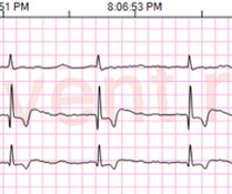
Patient had an unwitnessed cardiacarrest without bystander CPR performed. Initial vitals show hypertension (175/85), Atrial Fibrillation with RVR as seen in Figure 1 , hypercapnia (99mmHg), and SPO2 of 100%. Figure 2 : This rhythm shows a sinus bradycardia at a rate between 30 and 40bpm.
There is also bradycardia. Bradycardia puts patients at risk for "pause-dependent" Torsades de Pointes. Torsades in acquired long QT is much more likely in bradycardia because the QT interval following a long pause is longer still. If cardiacarrest from hypokalemia is imminent (i.e., mEq/L, from 1.9
Written by Magnus Nossen The patient in today's case is a male in his 70s with hypertension and type II diabetes mellitus. This patient is actively dying from a left main coronary artery OMI and cardiacarrest from VT/VF or PEA is imminent! Complete LMCA occlusion is associated with clinical shock and/or cardiacarrest.
Written by Jesse McLaren An 80 year old patient with diabetes/hypertension/ cirrhosis had a recent increase in candesartan for their hypertension, and was also on spirolactone and nadolol. Theres no prior ECG to compare - but the bradycardia, prolonged PR and peaked T waves could all be from hyperkalemia.
We organize all of the trending information in your field so you don't have to. Join thousands of users and stay up to date on the latest articles your peers are reading.
You know about us, now we want to get to know you!
Let's personalize your content
Let's get even more personalized
We recognize your account from another site in our network, please click 'Send Email' below to continue with verifying your account and setting a password.


Let's personalize your content