This site uses cookies to improve your experience. To help us insure we adhere to various privacy regulations, please select your country/region of residence. If you do not select a country, we will assume you are from the United States. Select your Cookie Settings or view our Privacy Policy and Terms of Use.
Cookie Settings
Cookies and similar technologies are used on this website for proper function of the website, for tracking performance analytics and for marketing purposes. We and some of our third-party providers may use cookie data for various purposes. Please review the cookie settings below and choose your preference.
Used for the proper function of the website
Used for monitoring website traffic and interactions
Cookie Settings
Cookies and similar technologies are used on this website for proper function of the website, for tracking performance analytics and for marketing purposes. We and some of our third-party providers may use cookie data for various purposes. Please review the cookie settings below and choose your preference.
Strictly Necessary: Used for the proper function of the website
Performance/Analytics: Used for monitoring website traffic and interactions
During the night, while on telemetry, the patient became bradycardic, with periods of isorhythmic AV dissociation (nodal escape rhythm alternating with sinus bradycardia), and there were sporadic PVCs. Cardiacarrest was called and advanced life support was undertaken for this patient. Without an MRI, it is impossible to know.
See our other countless hyperkalemia cases below: General hyperkalemia cases: A 50s year old man with lightheadedness and bradycardia Patient with Dyspnea. A woman with near-syncope, bradycardia, and hypotension What happens if you do not recognize this ECG instantly? HyperKalemia with CardiacArrest. With a twist.
While on telemetry monitoring he suffered cardiacarrest and was resuscitated. What ECG finding may have contributed to (or precipitated) the cardiacarrest? Learning points : Takotsubo can lead to cardiacarrest from ventricular arrhythmia. There are no clear signs of OMI. There is a prolonged QTc.
In-hospital cardiacarrest (IHCA) is a major healthcare problem with a high mortality rate. With continuous telemetry monitoring, heart rate trends could be used to predict IHCA events.
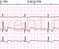
Patient had an unwitnessed cardiacarrest without bystander CPR performed. Crew notifies the received ED of an incoming post-arrest patient and notes a sinus bradycardia on their monitor, as seen in Figure 2. Figure 2 : This rhythm shows a sinus bradycardia at a rate between 30 and 40bpm.
Krantz et al authored a State-of-the-Art Review on Cardiovascular Complications of Opioid Use ( JACC 77(2):205-223, 2021 ) — in which mechanisms from Opioid Overdose that detail arrhythmia production ( up to cardiacarrest ) are elucidated — thereby providing an explanation for the unusual arrhythmias in today's case.
A 60-something woman presented after a witnessed cardiacarrest. This is commonly found after epinephrine for cardiacarrest, but could have been pre-existing and a possible contributing factor to cardiacarrest. Final Diagnosis: CardiacArrest due to Torsades from long QT of unknown etiology.
Discontinue all negative chronotropic agents, since the risk of torsade is much higher with bradycardia or pauses. See this post: How a pause can cause cardiacarrest 2. There is ventricular bigeminy with bizarre appearing wide T-waves See even more striking cases of this at the bottom of the post. The plan: 1.
Altered Mental Status, Bradycardia == MY Comment , by K EN G RAUER, MD ( 2/2 /2024 ): == Dr. Meyers began today’s case with the clinical challenge of asking you to identify the underlying cause of ECG #2. -- Read this ECG -- Osborn Waves and Hypothermia (this is the "Figure" above) What does LBBB look like in severe hypothermia?
One hour later (labs not yet returned), here is the ECG recorded just after the team noticed a sudden wide complex with precipitous decompensation, just before cardiacarrest: Bizarre, Brady, and Broad (wide QRS). Unfortunately, this was not recognized at this time. I believe it was this point when hyperkalemia was first suspected.
Uncontrolled coronary spasm may be associated with serious arrhythmias , including cardiacarrest ( Looi et al — Postgrad Med, 2012 ; Tan et al — Eur Heart J Case Rep, 2018 ; Chevalier et al — JACC, 1998 ; Rodriguez-Manero — EP Europace, 2018 ). Initial high sensitivity troponin I returned at 6ng/L (normal 0.20
This false electrical capture may have made cardiacarrest recognition difficult, and the re-arrest may have gone unrecognized for an unknown amount of time. Learning points: TCP is primarily recommended for bradycardia that does not respond to atropine, or other agents. Current 85mA. On ED arrival ROSC is achieved.
Her vital signs were within normal limits except for bradycardia at 55 bpm. It is probably sinus bradycardia with very small/depressed P-waves and prolonged PR interval. See these other related cases: A patient with cardiacarrest, ROSC, and right bundle branch block (RBBB). Is this just right bundle branch block?
Decades ago, an association of J waves with cardiacarrest (CA) or ventricular fibrillation (VF) was reported (1), whereas J waves have been observed in individuals in the general population.
There was no evidence bradycardia leading up to the runs of PMVT ( as tends to occur with Torsades ). If there had been — a temporary atrial pacemaker could have been considered as a way of increasing the heart rate to suppress a bradycardia-dependent arrhythmia ("overdrive pacing").
Polymorphic Ventricular Tachycardia Long QT Syndrome with Continuously Recurrent Polymorphic VT: Management CardiacArrest. A New Seizure in a Healthy 20-something More cases of long QT not measured correctly by computer (these are all fascinating ECGs/cases): Bupropion Overdose Followed by CardiacArrest and, Later, ST Elevation.
This patient is actively dying from a left main coronary artery OMI and cardiacarrest from VT/VF or PEA is imminent! Complete LMCA occlusion is associated with clinical shock and/or cardiacarrest. The arterial blood gas showed a lactic acidosis with a lactate level of 17mmol/L.
There is also bradycardia. Bradycardia puts patients at risk for "pause-dependent" Torsades de Pointes. Torsades in acquired long QT is much more likely in bradycardia because the QT interval following a long pause is longer still. If cardiacarrest from hypokalemia is imminent (i.e., mEq/L, from 1.9
Hyperkalemia causes peaked T waves and the "killer B's of hyperkalemia", including bradycardia, broad QRS complexes, blocks of the AV node and bundle branches, Brugada morphology, and otherwise bizarre morphology including sine wave. Steve, what do you think of this ECG in this CardiacArrest Patient?" With a twist.
Osborn waves have been reported with hypercalcemia, brain injury, subarachnoid hemorrhage, Brugada syndrome, cardiacarrest from VFib — and — severe, acute ischemia resulting in acute MI ( See My Comment in the November 22, 2019 post on Dr. Smith’s Blog ). Rituparna et al — as well as Chauhan and Brahma ( Int.
A fast heartbeat is called tachycardia, while a slow heartbeat is called bradycardia in medical terms. CardiacarrestCardiacarrest is a medical emergency in which the heart stops pumping blood to the body. Electrocardiogram, echocardiogram, and some other tests are done for patients with cardiacarrest.
Similarly, you may use our , app to adjust the paper speed along with amplification to read the slightest changes, especially for conditions like tachycardia and bradycardia. AI recognizing cardiacarrests in emergency calls. AI recognizing cardiacarrests in emergency calls.
That said — obvious findings include: i ) Marked bradycardia! — Unfortunately, before this could be accomplished — the patient went into cardiacarrest. She was successfully resuscitated — with a post-arrest rhythm similar to that seen in Figure-1. Cardiac cath did not reveal significant coronary disease!
Smith: This bizarre ECG looks like a post cardiacarrest ECG with probable acidosis or hyperkalemia in addition to OMI. The patient died of cardiogenic shock within 24 hours despite mechanical circulatory support. Below the J-point is marked out showing the ST pathologic deviations. What was the pH and K? Potassium 4,6.
There is sinus bradycardia with one PVC. There is "Shark Fin morphology" I saw this and thought for certain that this was going to be an LAD or left main occlusion as etiology of arrest, and etiology of profound ST Elevation in I, II, aVL, and V3-V6, and ST depression in III, V1 and V2. She then had a 12-lead: What do you think?
For example — marked bradycardia with unusual forms of advanced AV block — or marked bradycardia without evident P waves — or marked QRS widening with "shapeless" morphology — are all ECG indication of the need for immediate IV calcium until improvement of these ECG patterns.
Laddergram Illustration: The mechanism of AV block in ECG #2 is complex. I suspect there is dual-level Wenckebach out of the AV Node — as shown by my proposed laddergram that I've drawn in Figure-3 ( For more on the mechanism of dual-level AV block — CLICK HERE ).
In any case, there is bradycardia. First, what kind of arrest was this? It was a PEA or bradyasystolic arrest , not a shockable rhythm. Although most cardiacarrest from MI is due to ventricular fibrillation, some is due to high grade AV block, and so this could indeed be due to large acute STEMI. What is going on?
This ECG pattern may be diagnostic of B rugada S yndrome IF seen in association with: i ) a history of cardiacarrest; polymorphic VT; or of non-vagal syncope; and / or ii ) a positive family history of sudden death at an early age; and / or iii ) a similar ECG in relatives. Bradycardia. Acute febrile illness. Hypothermia.
U waves may also be found in patients with LVH and/or bradycardia , or occasionally as a normal variant. Low body magnesium is often encountered in association with other electrolyte abnormalities ( ie, low sodium, potassium, calcium or phosphorus ) ; acute MI; cardiacarrest; digoxin/diuretic use; alcohol use and abuse; renal impairment.
About two hours after admission, he suffered a cardiacarrest (whether it was VF/VT or PEA is not available) and expired. The provider contacted cardiology to discuss the case, but cardiology "didn't think it was a STEMI, didn't think he needed emergent cath." He was admitted to the cardiology floor and diagnosed with an NSTEMI.
In addition to a spontaneous or induced Brugada-1 ECG pattern, criteria for B rugada S yndrome require one or more of the following: History of cardiacarrest, of polymorphoic VT, or of non-vagal syncope — positive family history of sudden death at an early age — a similar ECG in close relatives.
These include ( among others ) — acute febrile illness — variations in autonomic tone — hypothermia — ischemia-infarction — malignant arrhythmias — cardiacarrest — and especially Hyperkalemia. Other Arrhythmias ( PACs, PVCs, AFib, Bradycardia and AV conduction disorders — potentially lethal VT/VFib ).
If a patient presents with chest pain and a normal heart rate, or with shockable cardiacarrest, then ischemic appearing ST elevation is STEMI until proven otherwise. CLICK HERE — for the ESC/ACC/AHA/WHF 2018 Consensus Document on the 4th Universal Definition of MI, in which these concepts are discussed and illustrated.
Written by Pendell Meyers, with edits by Steve Smith Thanks to my attending Nic Thompson who superbly led this resuscitation We received a call that a middle aged male in cardiacarrest was 5 minutes out. He was estimated to be in his 50s, with no known PMHx. He arrived with chest compressions ongoing, intubated, and being bagged.
Further history later: This patient personally has no further high risk features (syncope / presyncope), but her mother had sudden cardiacarrest in sleep. Regardless of further evaluation, she should avoid bradycardia, AV nodal blockers, Na channel blockers, and fevers. --If
This ECG shows a sinus bradycardia with a normal conduction pattern (normal PR, normal QRS, and normal QTc), normal axis, normal R-wave progression, normal voltages. Hypothermia can also produce bradycardia and J waves, with a pseudo-STEMI pattern. There is marked sinus bradycardia. What do you think? As per Drs.
Theres sinus bradycardia, borderline PR interval, narrow QRS; normal axis/R wave progression; low precordial voltages, and subtle peaked T waves (most obvious in V2, but all T waves are symmetric with a narrow base). Theres no prior ECG to compare - but the bradycardia, prolonged PR and peaked T waves could all be from hyperkalemia.
The patient was unconscious BEFORE the cardiacarrest, at the same time that she had strong pulses. Therefore, cardiacarrest is NOT the etiology of the coma. More cases here to highlight: [link] Middle Aged Woman with Asystolic CardiacArrest, Resuscitated: Cath Lab? OMI is a clinical diagnosis.
In just 90 minutes from presentation, the patient progressed from that very subtle ECG to cardiacarrest. Discussion: This is a case of an initial ECG showing very subtle signs of hyperkalemia. Dr. McLaren recently wrote an excellent blog post on a similar case.
We organize all of the trending information in your field so you don't have to. Join thousands of users and stay up to date on the latest articles your peers are reading.
You know about us, now we want to get to know you!
Let's personalize your content
Let's get even more personalized
We recognize your account from another site in our network, please click 'Send Email' below to continue with verifying your account and setting a password.


Let's personalize your content