This site uses cookies to improve your experience. To help us insure we adhere to various privacy regulations, please select your country/region of residence. If you do not select a country, we will assume you are from the United States. Select your Cookie Settings or view our Privacy Policy and Terms of Use.
Cookie Settings
Cookies and similar technologies are used on this website for proper function of the website, for tracking performance analytics and for marketing purposes. We and some of our third-party providers may use cookie data for various purposes. Please review the cookie settings below and choose your preference.
Used for the proper function of the website
Used for monitoring website traffic and interactions
Cookie Settings
Cookies and similar technologies are used on this website for proper function of the website, for tracking performance analytics and for marketing purposes. We and some of our third-party providers may use cookie data for various purposes. Please review the cookie settings below and choose your preference.
Strictly Necessary: Used for the proper function of the website
Performance/Analytics: Used for monitoring website traffic and interactions
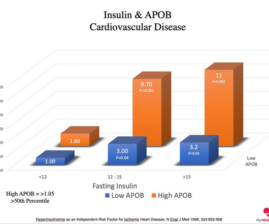
Excess weight, as measured by BMI, is typically considered a measure of excess body fat. Excess visceral fat results in a higher risk of insulin resistance, which is the precursor state to diabetes. In the setting of insulin resistance, a higher ApoB concentration increases the risk of cardiovascular disease dramatically.
Body mass index (BMI) is a widely available marker of nutrition status, however studies on BMI and post-ICH outcomes are limited and have conflicting results. Patients were divided into underweight, normal weight, overweight, and obese according to World Health Organization BMI criteria. 7.34) and lobar (OR 3.15, 95% CI 1.67-5.94)
Salvatore Carbone, PhD: First, I’d like to point out that obesity is a major risk factor for cardiometabolic disease. There are significant data that show that if you have obesity, you have a high risk of developing coronary heart disease, heart failure, type 2 diabetes (T2D) or risk factors such as hypertension and dyslipidemia. [1]
A, Normal axillary lymph nodes measuring milla1cf Fri, 05/10/2024 - 08:12 May 10, 2024 — According to the Summa Cum Laude Award-Winning Online Poster presented during the 124th ARRS Annual Meeting , fat-enlarged axillary nodes on screening mammograms can predict high cardiovascular disease (CVD) risk, Type 2 diabetes (T2DM), and hypertension (HTN).
An elevated Lp(a) is a common genetic factor that is independently and causally related to premature coronaryarterydisease. The occurrence of disease in this instance is probabilistic, not deterministic. An elevated Lp(a) does increase the risk of early cardiovascular disease, but that risk is not set in stone.
Short sleep duration was significantly associated with a higher risk of developing hypertension after adjusting for demographic and cardiovascular risk factors, including age, sex, education, BMI, blood pressure, smoking status etc. Sleep apnea has been tied to higher rates of high blood pressure, stroke and coronaryarterydisease.
However, recent studies have observed that people below 40 are also experiencing heart attacks due to high blood pressure, cholesterol, diabetes, smoking, obesity, a sedentary lifestyle, and social and mental stress. It’s essential for those at risk of coronaryarterydisease to be aware of the following symptoms.
Studies have shown that an increased left ventricular end-diastolic diameter (LVEDD) is associated with worse outcomes in various cardiovascular conditions, including heart failure, and coronaryarterydisease (CAD). The restrictive cubic spline in Figure 2 showed that LVEDD greater than 60 mm increased the risk of MACEs.
Diabetes, Red blood cell distribution width (RDW), and Triglyceride glucose-body mass index (TyG-BMI), as determined by Lasso regression and multivariate logistic regression.
Further regression analysis indicated that body mass index (BMI) might be related to changes in LAD. Additionally, the use of digoxin could affect changes in left ventricular ejection fraction.
We organize all of the trending information in your field so you don't have to. Join thousands of users and stay up to date on the latest articles your peers are reading.
You know about us, now we want to get to know you!
Let's personalize your content
Let's get even more personalized
We recognize your account from another site in our network, please click 'Send Email' below to continue with verifying your account and setting a password.


Let's personalize your content