This site uses cookies to improve your experience. To help us insure we adhere to various privacy regulations, please select your country/region of residence. If you do not select a country, we will assume you are from the United States. Select your Cookie Settings or view our Privacy Policy and Terms of Use.
Cookie Settings
Cookies and similar technologies are used on this website for proper function of the website, for tracking performance analytics and for marketing purposes. We and some of our third-party providers may use cookie data for various purposes. Please review the cookie settings below and choose your preference.
Used for the proper function of the website
Used for monitoring website traffic and interactions
Cookie Settings
Cookies and similar technologies are used on this website for proper function of the website, for tracking performance analytics and for marketing purposes. We and some of our third-party providers may use cookie data for various purposes. Please review the cookie settings below and choose your preference.
Strictly Necessary: Used for the proper function of the website
Performance/Analytics: Used for monitoring website traffic and interactions
You cannot eliminate the plaque entirely, but multiple clinical trials have shown plaque regression using high-intensity cholesterol-lowering treatments, which I have discussed previously. All of these parameters are important and need to be considered when evaluating plaque regression. REVERSAL Investigators.
The scan also showed “scattered coronary artery plaques”. __ Smith comment 1 : the appropriate management at this point is to lower the bloodpressure (lower afterload, which increases myocardial oxygen demand). The patient was put on a nitroglycerin drip and his pain improved with his bloodpressure.
The incidence of no-reflow was higher in patients with attenuated plaque ≥5 mm in length as evaluated by intravascular ultrasound (IVUS).Objective:The Bloodpressure decrease during PCI was significantly more pronounced in the no-reflow group (47.4% vs. 8.6%, p < 0.001). vs. 25.5%, p = 0.032). vs. 41.2%, p=0.043).Conclusion:In
Plaque regression can be demonstrated by ultrasound evaluation of the carotids which are easily accessible. Regular exercise can bring down the bloodpressure in the long run. Though bloodpressure rises progressively with increasing exercise, it reduces the resting bloodpressure in the long run.
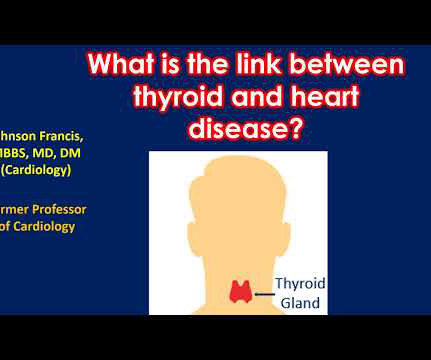
This in turn can enhance the chance of plaque build-up in the blood vessels of the heart (coronary arteries). If it is severe enough to compress the heart, it prevents proper filling of the heart and bloodpressure falls. Reduced function of the thyroid gland is also associated with heart disease.
ET Murphy Ballroom 4 Comparison of an "Inclisiran First" Strategy with Usual Care in Patients With Atherosclerotic Cardiovascular Disease: Results From the VICTORION-INITIATE Randomized Trial Targeting Weight Loss to Personalize the Prevention of Type 2 Diabetes Once-weekly Semaglutide in Patients with Heart Failure With Preserved Ejection Fraction, (..)
EMS obtained the following vital signs: pulse 50, respiratory rate 16, bloodpressure 96/49. Two thirds of MINOCA cases are due to atherosclerotic causes One way to prove the diagnosis in this case would have been with intravascular imaging such as optical coherence tomography (OCT) or intravascular ultrasound (IVUS).
Although it is statistically unlikely, multiple plaque ruptures are possible. On intravascular ultrasound (IVUS), the mid RCA plaque was described as "cratered, inflamed, and bulky," and the OM plaque was described as "bulky with evidence of inflammation and probably ulceration." Heitner et al.
At baseline visit, women will be randomized to undergo 2D/3D/strain vascular ultrasound (360 with imaging vs. 360 age- and RF-matched controls without imaging). Imaging can play a key role by revealing the presence of atherosclerotic plaques in a directly relatable way and thus, larger effects are anticipated in women with plaques.
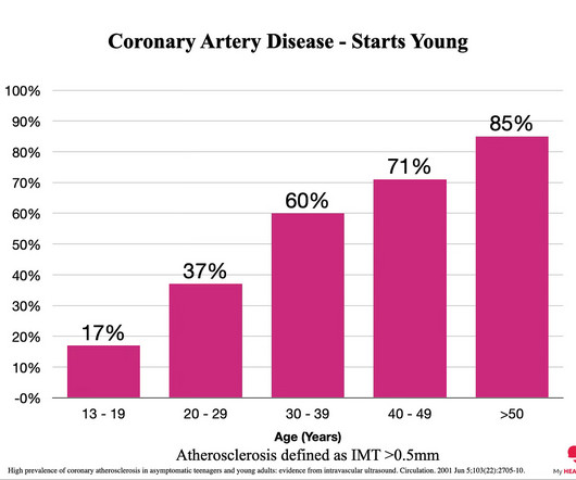
Everyone starts with no plaque in the coronary arteries, but over a long enough time frame, everyone develops plaque in their coronary arteries. By age 80, almost everyone will have evidence of advanced plaque in their coronary arteries, as defined by a cardiac CT 1. Plaque accumulation happens in stages. You got it.
Infections and inflammation of the heart eg myocarditis will cause acute inflammation of the heart and therefore may compromise the pumping ability of the heart Conditions such as high bloodpressure will make the heart work harder and as it does so it will become more muscular. The plaques can damage us in 2 ways.
They did not have an ultrasound on the ambulance (some local crews are starting to utilize POC limited US in our service areas). He was taken to the cath lab and underwent emergent intervention: Thrombotic stenosis of the proximal RCA (95% with evidence of plaque rupture) is the culprit for the patient's inferoposterior STEMI.
Heart disease, the build-up of plaque in the coronary arteries, typically starts years, if not decades, prior to an event. These noninvasive scans look directly at the coronary arteries rather than assessing for the risk factors for coronary artery disease eg LDL cholesterol, high bloodpressure etc. CT Coronary Angiogram.
The bloodpressure was 170/100 in the critical care area. Cardiology wanted a CT of the aorta to rule out dissection, presumably partly due to the very high bloodpressure readings, but also because it is hard for people to believe that a 20-something woman could have acute thrombotic coronary artery. Ultrasound Med.
We organize all of the trending information in your field so you don't have to. Join thousands of users and stay up to date on the latest articles your peers are reading.
You know about us, now we want to get to know you!
Let's personalize your content
Let's get even more personalized
We recognize your account from another site in our network, please click 'Send Email' below to continue with verifying your account and setting a password.


Let's personalize your content