This site uses cookies to improve your experience. To help us insure we adhere to various privacy regulations, please select your country/region of residence. If you do not select a country, we will assume you are from the United States. Select your Cookie Settings or view our Privacy Policy and Terms of Use.
Cookie Settings
Cookies and similar technologies are used on this website for proper function of the website, for tracking performance analytics and for marketing purposes. We and some of our third-party providers may use cookie data for various purposes. Please review the cookie settings below and choose your preference.
Used for the proper function of the website
Used for monitoring website traffic and interactions
Cookie Settings
Cookies and similar technologies are used on this website for proper function of the website, for tracking performance analytics and for marketing purposes. We and some of our third-party providers may use cookie data for various purposes. Please review the cookie settings below and choose your preference.
Strictly Necessary: Used for the proper function of the website
Performance/Analytics: Used for monitoring website traffic and interactions
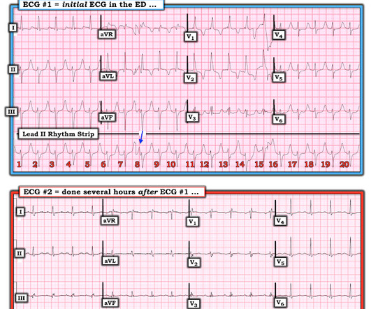
This EKG is diagnostic of transmural ischemia of the inferior wall. Smith: note also the terminal QRS distortion in lead III (absence of S-wave without a prominent J-wave). __ Smith comment 1 : the appropriate management at this point is to lower the bloodpressure (lower afterload, which increases myocardial oxygen demand).
BackgroundAcute myocardial ischemia (AMI)triggered ventricular arrhythmias are closely linked to maladaptive sympathetic hyperactivity mediated via the left stellate ganglion (LSG). Retigabine pretreatment significantly suppressed ischemiainduced LSG hyperactivity and reduced sympathetic activation markers compared with controls.
BACKGROUND:Abnormal orthostatic bloodpressure (BP) regulation may result in cerebral hypoperfusion and brain ischemia and contribute to dementia. Hypertension, Ahead of Print. It may also manifest as early symptoms of the neurodegenerative process associated with dementia.
The initial bloodpressure was 80/palp with a heart rate of 104, respirations 20, oxygen saturations of 94% and a finger stick blood glucose of 268. In addition, the patient received 750 mL of fluid resuscitation with transient improvement of bloodpressure.
Maternal blood and tissues were collected. Bloodpressure increased in RUPP rats and was normalized with rituximab treatment. A separate group of dams were allowed to deliver, pup weights were recorded, and at 4 months of age, tissues were collected from offspring.
This may be in the strength of the pulse ( or the bloodpressure recorded ) — or it may be in one or more waveforms in the ECG recording. Alternation in ST segment appearance ( or in the amount of ST elevation or depression ) — is often linked to ischemia.
ObjectiveA significant proportion (85%) of low-risk non-ST-elevation acute coronary syndrome (NSTE-ACS) patients do not receive objective confirmation of ischemia by stress echocardiography (SE), yet remain a healthcare burden due to lower long-term survival and overuse of medical services.
ET Main Tent (Hall B1) This session offers more insights from key clinical trials presented at ACC.24 24 and find out what it all means for your patients.
Are you confident there is no ischemia? Primary VT , and the VT with tachycardia is causing ischemia with chest discomfort (supply-demand mismatch/type 2 MI)? Ischemia from ACS causing the chest discomfort, with VT another consequence (or coincidence)? Do you agree with this strategy? How can you better assess the ST segments?
This proves effective treatment of the recurrent ischemia. The patient had no further symptoms of ischemia. EKG 3 is diagnostic for developing re-occlusion, and EKG 4 proves that the nitrates relieved the ischemia. = This proves effective treatment of the recurrent ischemia." Here was her final EKG prior to discharge.
Contrary to what Ken stated, the ST vector remains mostly posterior __ What about subendocardial ischemia? Subendocardial ischemia results in ST depression, but unfortunately, and rather mysteriously, it does not localize to the ischemic wall. Similarly, STD in aVL is usually reciprocal to inferior ST elevation, not "lateral ischemia."
Vascular inflammation underlies the development of hypertension, and the mechanisms by which it increases bloodpressure remain the topic of intense investigation. Hypertension, Ahead of Print. These pathways share a dependence upon redox signaling, and excessive activation promotes oxidative stress that promotes vascular aging.
Though this association may be related to impaired cerebral oxygen delivery, it is unclear whether these changes relate to cerebral ischemia. Background:We have previously identified that hemoglobin decrements and new-onset anemia during an intracerebral hemorrhage (ICH) hospitalization is frequent, rapid, and associates with poor outcome.
He presented to the Emergency Department with a bloodpressure of 111/66 and a pulse of 117. One very useful adjunct is ultrasound: Echo of his heart can distinguish aneurysm from acute MI by presence of diastolic dyskinesis, but it cannot distinguish demand ischemia from ACS. He had this ECG recorded.
Introduction:Oxidative stress plays an important role in both early brain injury and delayed cerebral ischemia after subarachnoid hemorrhage (SAH). There were no differences in bloodpressure or body weight between the two groups. Stroke, Volume 56, Issue Suppl_1 , Page AWP368-AWP368, February 1, 2025. P<0.05, Fig.
Bloodpressure was 150/80. He had no previous history of CAD, and presented with very typical waxing and waning chest pain, much worse with exertion but also present at rest and on presentation, though his pain was minimal at the time of the ECG. This is all suggestive of posterior STEMI, but not definitely diagnostic.
Acute PE can also result in right precordial ST elevation due to RV stress and ischemia ) 90 minutes later, I was shown this followup ECG without any knowledge of the ultrasound results: There is significantly less ST elevation, and the ST segment is now concave. The RV was small and IVC empty, making pulmonary embolism extremely unlikely.
Pericardial tamponade is also associated with pulsus paradoxus which is an abnormally large drop in systolic bloodpressure greater than 10 mmHg during inspiration. This may be in the strength of the pulse ( or the bloodpressure recorded ) — or it may be in one or more waveforms in the ECG recording.
The ECG was incorrectly interpreted as no signs of ischemia. He is placed on heparin drip, he will have IV beta-blocker and oral beta blocker for heart rate control and bloodpressure management. A prior ECG was available: This ECG is normal, and thus confirms the concerns explained above.
Bloodpressure was normal (109/83). NOTE #3: In the context of a long QTc or ischemia — the finding of ST segment and/or T wave alternans may predict the occurrence of malignant ventricular arrhythmias. A 30-something was in the ED for some minor trauma when he was noted to have a fast heart rate. but only when asked.
Either could be a result of myocardial contusion There is some minimal ST depression -- this could represent ischemia What else is there that could use therapy immediately? Or, much less likely, it could be a very accelerated escape rhythm from the posterior fascicle. There is a very long ST segment resulting in a very long QT. mEq/L and 3.8
The flutter waves can conceal or mimic ischemic repolarization findings, but here I don't see any obvious findings of OMI or subendocardial ischemia. 2) Norepinephine to support BloodPressure. The rhythm is 2:1 atrial flutter. If this were RBBB, it would need to be incomplete, as the QRS duration is less than 120 ms.
High BloodPressure (Hypertension) Persistent high bloodpressure forces the heart to work harder to pump blood. Coronary Artery Disease (CAD) CAD, which involves the narrowing or blockage of coronary arteries due to plaque buildup, can reduce blood flow to the heart. Anticoagulants to prevent blood clots.
The first task when assessing a wide complex QRS for ischemia is to identify the end of the QRS. The ST segment changes are compatible with severe subendocardial ischemia which can be caused by type I MI from ACS or potentially from type II MI (non-obstructive coronary artery disease with supply/demand mismatch). What do you think?
BackgroundDelayed cerebral ischemia represents a significant contributor to death and disability following aneurysmal subarachnoid hemorrhage. Journal of the American Heart Association, Ahead of Print. Our analysis included 102 eligible studies. Vasospastic events were mainly assessed through microscopy of large cerebral arteries.
Risk factors for PAD include smoking; having Type 1 or Type 2 diabetes, high bloodpressure, high cholesterol, chronic kidney disease, atherosclerosis in other parts of the body (such as coronary artery disease); and being age 75 years or older. and Global Data From the American Heart Association.
The relationship of cSS with DWI lesion presence as the outcome was assessed with logistic regression adjusted for ICH score, bloodpressure change within 24 hours, and small vessel disease markers (CMB and white matter hyperintensity). DWI hyperintensities were identified if >10mm from the hematoma with decreased ADC signal.
There is appreciable STE aVR with near-global STD that appropriately maximizes in Leads II and V5, and thus suggesting a circumstance of generic, diffusely populated, circumferential subendocardial ischemia versus occlusive coronary thrombus. [1] Although the bloodpressure resolved, his pain, however, did not.
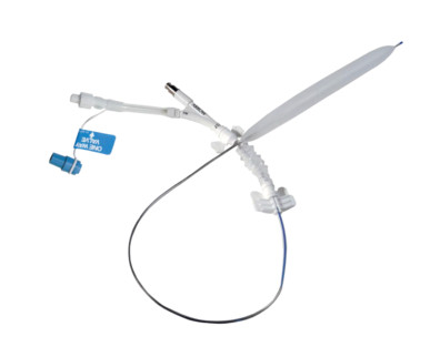
This issue may: prevent the balloon from fully inflating, cause blood to back up in the tubing, allow helium to leak, and lead to catheter damage or insertion difficulty during use. Teleflex/Arrow International reports 322 complaints. A total of 31 injuries and 3 deaths have been reported potentially related to this issue.
Due to conflicting prior studies, it is uncertain if the degree of systolic bloodpressure reduction increases the risk. Patients with severe, chronic hypertension may be more vulnerable to the development of ischemia after ICH due to altered cerebral autoregulatory limits. and LVH was seen in 23.5%.
When I was shown this ECG, I said it looks like such widespread ischemia that is might be a left main occlusion, or LM ischemia plus circumflex occlusion (high lateral and posterior OMI). There is STE in aVR. Thus, there is high lateral OMI with diffuse ST depression. Moreover, left main occlusion often presents near death.
Immediately after contrast injection into the LMCA, the patient had circulatory collapse, with a precipitous drop in bloodpressure. An Impella device was placed to maintain cardiac output and perfusion pressures. There is no definite evidence of acute ischemia. (ie, Epinephrine infusion was begun.
In MSIMI (Mental Stress-induced Myocardial Ischemia) studies , mental stress activities like public speaking were evaluated for their impact on ischemia, measured via myocardial SPECT and vascular function (microvascular function, endothelial function).
This ECG is diagnostic of diffuse subendocardial ischemia. The combination of sudden increased intracranial pressure with loss of spontaneous circulation results in near total loss of cerebral perfusion. The bloodpressure produced by chest compressions is inadequate to perfuse the brain when ICP is high. Bart BA.
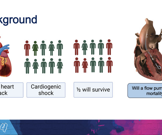
However, the results also showed significantly higher rates of complications among patients who received the heart pump, including bleeding, limb ischemia, renal replacement therapy and sepsis. “It It doesn’t come without a cost—we see significantly more serious complications in the Impella treated patients,” Møller said.
RCA ischemia often results in sinus bradycardia from vagal reflex or ischemia of the sinus node. During transport, I had debated giving atropine for his bradycardia and cardiogenic shock, but was worried about making an already profoundly ischemic heart more ischemic, and chose instead to optimize preload with pressure bagging 1L NS.
1,12,13 While it is important to treat all known risk factors that contribute to ASCVD including high bloodpressure, hyperlipidemia, diabetes, and obesity, physicians also need to recognize and treat systemic inflammation in CV disease. This in turn leads to an overall reduction in IL-6 production and CRP concentration.12
Because the evidence suggests that when we do, patients end up with better bloodpressure, cholesterol control and are more likely to engage in healthier behaviours. 5 ISCHEMIA Research Group. Historically, I used to describe the report and then draw a diagram, but I show patients their own scan results more and more.
The EMS narrative reports that her bloodpressure and oxygenation improved modestly with rhythm stability for transport duration. In most cases, rather, the culprit is gross ischemia due to myocardial infarction, cardiomyopathy, or advanced coronary artery disease. Unfortunately, a post-conversion 12 Lead was not acquired.
On arrival in the ED, he was hypotensive with a systolic bloodpressure in the 70s. These include ( among others ) — acute febrile illness — variations in autonomic tone — hypothermia — ischemia/infarction/cardiac arrest — and Hyperkalemia. Fluid resuscitation was initiated. Here is the initial ED ECG: What do you think?
Evidence of acute ischemia (may be subtle) vii. Any ED systolic bloodpressure less than 90 or greater than 180 mm Hg (+1) 4. The cost per test affecting diagnosis or management was highest for electroencephalography ($32,973), CT ($24,881), and cardiac enzymes ($22,397) and lowest for postural bloodpressure ($17-$20).
They were unable to obtain a bloodpressure. His heart rate was in the low 20s and we were also unable to obtain a bloodpressure. He was given 50 mcg epinephrine with good response in both heart rate and bloodpressure. There is Transmural ischemia of Occlusion MI. His temperature was 32.8
BackgroundPainful left bundle branch block (LBBB) syndrome is an uncommon disease that is defined as intermittent episodes of angina associated with simultaneous LBBB changes on an electrocardiogram (ECG) with the absence of flow-limiting coronary artery disease or ischemia on functional testing.
We organize all of the trending information in your field so you don't have to. Join thousands of users and stay up to date on the latest articles your peers are reading.
You know about us, now we want to get to know you!
Let's personalize your content
Let's get even more personalized
We recognize your account from another site in our network, please click 'Send Email' below to continue with verifying your account and setting a password.


Let's personalize your content