This site uses cookies to improve your experience. To help us insure we adhere to various privacy regulations, please select your country/region of residence. If you do not select a country, we will assume you are from the United States. Select your Cookie Settings or view our Privacy Policy and Terms of Use.
Cookie Settings
Cookies and similar technologies are used on this website for proper function of the website, for tracking performance analytics and for marketing purposes. We and some of our third-party providers may use cookie data for various purposes. Please review the cookie settings below and choose your preference.
Used for the proper function of the website
Used for monitoring website traffic and interactions
Cookie Settings
Cookies and similar technologies are used on this website for proper function of the website, for tracking performance analytics and for marketing purposes. We and some of our third-party providers may use cookie data for various purposes. Please review the cookie settings below and choose your preference.
Strictly Necessary: Used for the proper function of the website
Performance/Analytics: Used for monitoring website traffic and interactions
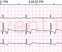
Patient had an unwitnessed cardiacarrest without bystander CPR performed. Crew notifies the received ED of an incoming post-arrest patient and notes a sinus bradycardia on their monitor, as seen in Figure 2. Figure 2 : This rhythm shows a sinus bradycardia at a rate between 30 and 40bpm.
Written by Pendell Meyers, with edits by Steve Smith Thanks to my attending Nic Thompson who superbly led this resuscitation We received a call that a middle aged male in cardiacarrest was 5 minutes out. He was estimated to be in his 50s, with no known PMHx. He arrived with chest compressions ongoing, intubated, and being bagged.
One hour later (labs not yet returned), here is the ECG recorded just after the team noticed a sudden wide complex with precipitous decompensation, just before cardiacarrest: Bizarre, Brady, and Broad (wide QRS). Compartment pressures in the right calf were all 40-50 mmHg. Unfortunately, this was not recognized at this time.
On arrival in the emergency department, invasive bloodpressure was 35/15mmHg and the patient was in profound cardiogenic shock with severe confusion secondary to brain hypoperfusion. The arterial blood gas showed a lactic acidosis with a lactate level of 17mmol/L. PUSH THE LYTICS ! The below ECG (ECG #4) was recorded.
Immediately after contrast injection into the LMCA, the patient had circulatory collapse, with a precipitous drop in bloodpressure. An Impella device was placed to maintain cardiac output and perfusion pressures. There was no evidence bradycardia leading up to the runs of PMVT ( as tends to occur with Torsades ).
On arrival in the ED, he was hypotensive with a systolic bloodpressure in the 70s. Hyperkalemia causes peaked T waves and the "killer B's of hyperkalemia", including bradycardia, broad QRS complexes, blocks of the AV node and bundle branches, Brugada morphology, and otherwise bizarre morphology including sine wave.
They were unable to obtain a bloodpressure. His heart rate was in the low 20s and we were also unable to obtain a bloodpressure. He was given 50 mcg epinephrine with good response in both heart rate and bloodpressure. His rhythm on telemetry seemed to be sinus bradycardia vs junctional rhythm.
His first recorded bloodpressure was 88/53 mm Hg. Forty five minutes later, his bloodpressure increased to 157/125 mm Hg, but his heart rate was now in the 30s. The next recorded bloodpressure was 211/175 mm Hg, and in response the patient was started on continuous nitroglycerin infusion.
We organize all of the trending information in your field so you don't have to. Join thousands of users and stay up to date on the latest articles your peers are reading.
You know about us, now we want to get to know you!
Let's personalize your content
Let's get even more personalized
We recognize your account from another site in our network, please click 'Send Email' below to continue with verifying your account and setting a password.


Let's personalize your content