This site uses cookies to improve your experience. To help us insure we adhere to various privacy regulations, please select your country/region of residence. If you do not select a country, we will assume you are from the United States. Select your Cookie Settings or view our Privacy Policy and Terms of Use.
Cookie Settings
Cookies and similar technologies are used on this website for proper function of the website, for tracking performance analytics and for marketing purposes. We and some of our third-party providers may use cookie data for various purposes. Please review the cookie settings below and choose your preference.
Used for the proper function of the website
Used for monitoring website traffic and interactions
Cookie Settings
Cookies and similar technologies are used on this website for proper function of the website, for tracking performance analytics and for marketing purposes. We and some of our third-party providers may use cookie data for various purposes. Please review the cookie settings below and choose your preference.
Strictly Necessary: Used for the proper function of the website
Performance/Analytics: Used for monitoring website traffic and interactions
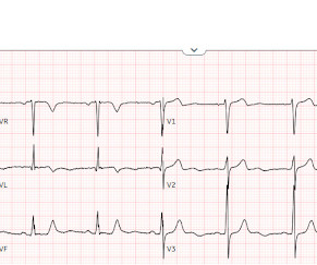
The patient with no prior cardiac history presented in the middle of the night with acute chest pain, and had this ECG recorded during active pain: I did not see any ischemia on this electrocardiogram. The patient has active chest pain, so if these are abnormally large T-waves This link shows 13 blog posts of Posterior Reperfusion T-waves.
See > 50 cases on Dr. Smiths EGC Blog. Weve also published the largest study on this question: Emergency Department Code STEMI patients with initial electrocardiogram labeled normal by computer interpretation: a 7-year retrospective review. Safety of computer interpretation of normal triage electrocardiograms. Take home 1.
Dr. Smith’s ECG Blog has published a growing list of over 40 cases of ECGs falsely labeled ‘normal’ by the computer which are diagnostic of Occlusion MI, and Smith et al. Smith’s ECG Blog has published a growing list of over 40 cases of ECGs falsely labeled ‘normal’ by the computer which are diagnostic of Occlusion MI, and Smith et al.
We have shown many examples of this on this blog. Emergency department Code STEMI patients with initial electrocardiogram labeled ‘normal’ by computer interpretation: a 7-year retrospective review. As we often emphasize on Dr. Smith's ECG Blog — there is normally slight, upward sloping ST elevation in leads V2 and V3.
This is because until recently — computerized programs have been based on STEMI -criteria — which as we have shown, will miss an estimated 25-30% of acute coronary occlusions ( See My Comment in the July 31, 2020 post in Dr. Smith's ECG Blog ). Am J Emerg Med. 2022 Jan;51:384-387. doi: 10.1016/j.ajem.2021.11.023. 2021.11.023.
The post 6 Cardiology Board Review Questions That Will Help You Pass the Boards appeared first on BoardVitals Blog. Question banks are a favorite exam preparation resource for Cardiologists that want to practice in the format of the exam. If you’d like more sample questions then follow this link to begin a free trial today.
International evaluation of an artificial-intelligence- powered electrocardiogram model detecting acute coronary occlusion myocardial infarction. Immediate and early percutaneous coronary intervention in very high-risk and high-risk non-ST segment elevation myocardial infarction patients. Clin Cardiol 2022 4. Herman, Meyers, Smith et al.
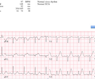
An initial electrocardiogram (ECG) is provided below. See My Comment at the bottom of the page in the May 19, 2020 post in Dr. Smith's Blog ). His current medication regimen includes apixaban, carvedilol, perindopril, spironolactone, torasemide, dapagliflozin, amiodarone, and ivabradine. What do you think? What is the rhythm?
This is obviously unreliable data, as Dr. Smith’s Blog has published 51 cases of OMI with ECGs labeled ‘normal’ , 35 of which were identified by the Queen of Hearts – with 10 examples here. Emergency department Code STEMI patients with initial electrocardiogram labeled ‘normal’ by computer interpretation: a 7-year retrospective review.
Because: 1) He has been reading this blog for a long time. The attending provider wrote “Agree with electrocardiogram interpretation”. Editorial Comment: I begin my thoughts on today's Blog post with the above reflections to provide perspective for my concerns about this case. 2) He is curious This is how Pendell got started.
Chest Pain Severity Rating Is a Poor Predictive Tool in the Diagnosis of ST-Segment Elevation Myocardial Infarction [link] Abstract Current ST-segment elevation myocardial infarction (STEMI) guidelines require persistent electrocardiogram ST-segment elevation, cardiac enzyme changes, and symptoms of myocardial ischemia.
can cause ST-segment elevation (STE) on electrocardiogram (ECG), the distinction between them may be hard and complicated. Many researchers, including the editors of this blog, tried to develop such tools in the recent past and we have recommended their use in certain clinical scenarios in many posts on this blog.
In the hope of dispelling continued dependence on millimeter-based STEMI criteria — we’ve published numerous cases in recent years in Dr. Smith’s ECG Blog of acute OMI ( O cclusion-based M yocardial I nfarction ) , in which patients have benefited from acute reperfusion despite not satisfying “STEMI criteria”.
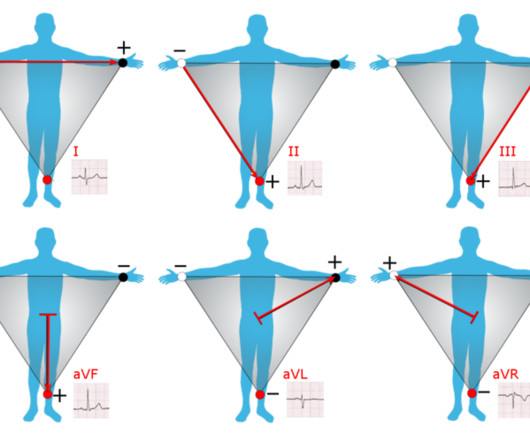
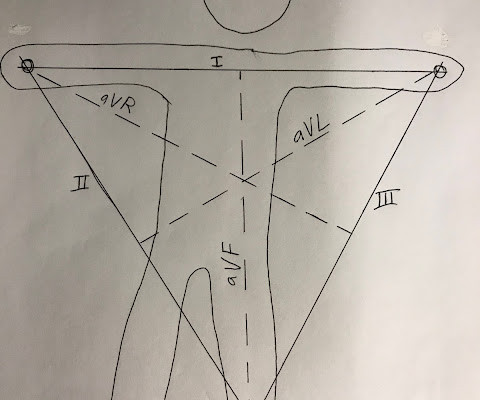
Introduction to ECG Testing & Einthoven’s Triangle: The electrocardiogram (ECG) represents the heart’s electrical activity, resulting from the contraction (depolarization) and relaxation (repolarization) of the atrial and ventricular muscles. Let’s start with grasping the basics of electrocardiography.
Usefulness of the Electrocardiogram in Establishing the Diagnosis and Prognosis of Arrhythmogenic Right Ventricular Cardiomyopathy Other References, from the above article: 1 FI Marcus Epsilon waves aid in the prognosis and risk stratification of patients with ARVC/D J Cardiovasc Electrophysiol, 26 (2015), pp. J Electrocardiol, 42 (2009), pp.
International evaluation of an artificial intelligence-powered electrocardiogram model detecting acute coronary occlusion myocardial infarction. Accuracy of OMI ECG findings versus STEMI criteria for diagnosis of acute coronary occlusion myocardial infarction. IJC Heart and Vasc 2021 8. Herman et al. Eur Herat J Digital Health 2024
That was a surprising finding, and it suggests that the CineECG could have more information than just a 12-lead electrocardiogram”… If you want to listen to the podcast here’s the link and if you just want to read the transcript here’s another link.
Explanation: Shown electrocardiogram suggests left ventricular hypertrophy. Shown electrocardiogram suggests left ventricular hypertrophy. The post Hypertropic Cardiomyopathy: A Board Review Question Explained By Video appeared first on BoardVitals Blog. Hypertrophic cardiomyopathy is one of them. Start with a Free Trial.
In this blog post, learn more about the advantages of a portable ECG device and how it can change how you track your heart health. Even if they are in a different place from the patient at the time of the test, medical professionals can quickly and conveniently check the electrocardiogram (ECG) results.
This blog explores how medical devices are becoming useful for patient compliance in areas like precision diagnostics, real-time monitoring, targeted therapy, and enhanced patient experience by personalizing medicines. These medical devices collect essential data, analyze information, and deliver personalized medicine.
Safety of Computer Interpretation of Normal Triage Electrocardiograms. The Comparison of Physician to Computer Interpreted Electrocardiograms on ST-elevation Myocardial Infarction Door-to-balloon Times. It is not yet available, but this is your way to get on the list. link] Hughes KE et al. Acad Emerg Med 2017; 24(1): 120 – 24.
D) An electrocardiogram is most commonly normal in these patients. D) An electrocardiogram is most commonly normal in these patients. appeared first on BoardVitals Blog. (A) This condition is characterized by transient regional systolic dysfunction of the left ventricle (LV). (B) What is Takotsubo Cardiomyopathy?
Below is the post -PCI electrocardiogram. For those in search of brief review of the Cabrera Format for ECG recording — Please check out My Comment at the bottom of the page in the October 26, 2020 post in Dr. Smith's ECG Blog. In the cath lab the patient was found to have a 100% occlusion of a small 1st marginal branch of the LCx.
Electromechanical association: a subtle electrocardiogram artifact. A very similar case to the one presented today appears in the January 17, 2023 post of Dr. Smith’s ECG Blog. Acute chest pain and a bizarre ECG Bizarre (Hyperacute??) T-waves More info on arterial pulse tapping artifact [link] Aslanger E, Yalin K. 2012;45(1):15-17.
Physician accuracy in interpreting potential ST-segment elevation myocardial infarction electrocardiograms. I believe there is not quite enough STE for formal STEMI criteria, but some might measure 1.0 Journal of the American Heart Association 2013;2:e000268. Carley et al. What’s the point of ST elevation? Emerg Med J. 2002;19:126-128.
The utility of the triage electrocardiogram for the detection of ST-segment elevation myocardial infarction. For those of you who read this blog regularly: You will know that these are clearly hyperacute T-waves , diagnostic of proximal LAD occlusion. This paper was just published: Noll S. Am J Emerg Med 36(10):1771-1774. October 2018.
Dedicated followers of the Smith ECG Blog know that the STD of true subendocardial ischemia does not localize, yet some of the examples listed below demonstrate the opposite, and were subsequently labeled “diffuse ischemia” or “generic subendocardial changes” as a diagnosis of convenience. However, the maximal STD in this case is V3.
Laurence Katz and Jonathan Jones Safety of Computer Interpretation of Normal Triage Electrocardiograms (pages 120–124). The Comparison of Physician to Computer Interpreted Electrocardiograms on ST-elevation Myocardial Infarction Door-to-balloon Times. This one was not even so subtle!!! References : 1. Hug hes KE., January 2017.
New insights into the use of the 12-lead electrocardiogram for diagnosing acute myocardial infarction in the emergency department. All electrocardiograms (ECGs) and coronary angiograms were blindly analyzed by experienced cardiologists. Harhash AA, Huang JJ, Reddy S, et al. aVR ST segment elevation: acute STEMI or not?
Updates on the Electrocardiogram in Acute Coronary Syndromes. Electrocardiogram patterns in acute left main coronary artery occlusion. Therefore, to be safe, an internal defibrillator was placed. The patient was discharged neurologically intact. References : 1. Current Emergency and Hospital Medicine Reports (2013) 1:4352.
A 12-lead electrocardiogram, lead V4R , and leads V7-9 were recorded on admission. OTHER Examples of Lead Reversal on Dr. Smith's Blog: Technical errors featuring a variety of lead reversal placements remain a surprisingly common “mishap” of everyday practice.
That said — followers of Dr. Smith's ECG Blog have already seen numerous clinical cases that we have presented in which the PM Cardio AI Bot app. Grauer K, Kravitz L, Curry RW, Ariet M: Computerized Electrocardiogram Interpretations: Are They Useful for the Family Physician? What About the Initial ECG? J Am Bd Fam Prac 1:17-24, 1989.
In Wung’s study, 81% of patients with greater than or equal to 1mm STE in posterior leads also had other significant STE on the 12-lead ECG, and 96% had some ST deviation. 3 However, 22-39% of patients experiencing posterior MI who have greater than or equal to 0.5mm STE in the posterior leads do not demonstrate STD in V1-V3.
At the bottom of the post, I have re-printed the section on aVR in my article on the ECG in ACS from the Canadian Journal of Cardiology: New Insights Into the Use of the 12-Lead Electrocardiogram for Diagnosing Acute Myocardial Infarction in the Emergency Department Case 1. Updates on the Electrocardiogram in Acute Coronary Syndromes.
Electromechanical association: a subtle electrocardiogram artifact. Arterial pulse tapping artifact [link] This online article references the article below by Emre Aslanger, a great guy who occasionally corresponds with me about ECGs. Aslanger E, Yalin K. Journal of Electrocardiology. 2012;45(1):15-17. doi:10.1016/j.jelectrocard.2010.12.162.
Below is from a quote from part of a piece on aVR which I wrote for Current Emergency and Hospital Medical Reports: " Updates on the electrocardiogram in Acute Coronary Syndromes. " Value of the electrocardiogram in localizing the occlusion site in the left anterior descending coronary artery in acute myocardial infarction.
Smith , d and Muzaffer Değertekin a DIFOCCULT: DIagnostic accuracy oF electrocardiogram for acute coronary OCClUsion resuLTing in myocardial infarction. His first electrocardiogram ( ECG) is given below: --Sinus bradycardia. Blood pressure: 130/80 mmHg, heart rate: 45/min, respiratory rate: 18/min, SaO2: %98, body temperature: normal.
Quote from the article: Since I do not know how to make a "degree" sign on this blog, I use "^" for degree sign : "Deviation of the mean electrical QRS axis of the heart in the frontal plane (A^QRS) from apparent normal values has been considered evidence of electrocardiographic abnormality. Journal of Electrocardiology 3(2):161-167; 1970.
A deep neural network for 12-lead electrocardiogram interpretation outperforms a conventional algorithm, and its physician over-read, in the diagnosis of atrial fibrillation. M Y A NSWER: The issue of whether C omputerized E CG I nterpretations are “at fault” for an inaccurate ECG diagnosis has been addressed numerous times on this blog.
Comment This paper has received some press recently: Safety of Computer Interpretation of Normal Triage Electrocardiograms The algorithm used was also the GE Marquette 12 SL. I don't know what her subsequent cardiac function was, but that is not the point of this post. But it could have been a disaster.
20 The primary objective of the Paced Electrocardiogram Requiring Fast Emergent Coronary Therapy (PERFECT) study was to compare the sensitivity and specificity of the MSC with those of the original Sgarbossa criteria for the diagnosis of occlusion myocardial infarction in patients with ventricular paced rhythm.
Traditional tools like stethoscopes, blood pressure gauges, and electrocardiograms (ECG) are fundamental for standard diagnostic practices. Medical Devices Both traditional and emerging medical devices play crucial roles in advancing cardiac diagnosis.
You will note that it is essentially an unremarkable electrocardiogram except for some PACS. We have often referred to this almost magical m irror- i mage r elationship for ST-T waves in leads I II and aV L — which when present, means a cute i nferior O MI until you prove otherwise ( See My Comment on the 8/9/2018 SSmith Blog Post ).
We organize all of the trending information in your field so you don't have to. Join thousands of users and stay up to date on the latest articles your peers are reading.
You know about us, now we want to get to know you!
Let's personalize your content
Let's get even more personalized
We recognize your account from another site in our network, please click 'Send Email' below to continue with verifying your account and setting a password.


Let's personalize your content