This site uses cookies to improve your experience. To help us insure we adhere to various privacy regulations, please select your country/region of residence. If you do not select a country, we will assume you are from the United States. Select your Cookie Settings or view our Privacy Policy and Terms of Use.
Cookie Settings
Cookies and similar technologies are used on this website for proper function of the website, for tracking performance analytics and for marketing purposes. We and some of our third-party providers may use cookie data for various purposes. Please review the cookie settings below and choose your preference.
Used for the proper function of the website
Used for monitoring website traffic and interactions
Cookie Settings
Cookies and similar technologies are used on this website for proper function of the website, for tracking performance analytics and for marketing purposes. We and some of our third-party providers may use cookie data for various purposes. Please review the cookie settings below and choose your preference.
Strictly Necessary: Used for the proper function of the website
Performance/Analytics: Used for monitoring website traffic and interactions
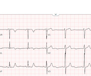
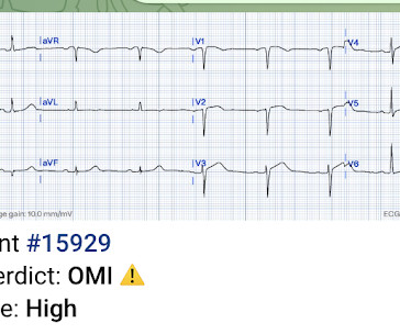
2 middle aged males presented with chestpain. Which had the more severe chestpain at the time of the ECG? Patient 2 at the bottom with a very subtle OMI complained of 10/10 chestpain at the time the ECG was recorded. 414 patients were included in the analysis.
Written by Jesse McLaren, with a very few edits by Smith A 60-year-old presented with chestpain. Inferior hyperacute T waves, which have been added to the 2022 ACC consensus on chestpain as a “STEMI equivalent”[3] 3. But are there any other signs of Occlusion MI? Conduction disorders in the setting of acute STEMI.
Written by Jesse McLaren Four patients presented with chestpain. Dr. Smith’s ECG Blog has published a growing list of over 40 cases of ECGs falsely labeled ‘normal’ by the computer which are diagnostic of Occlusion MI, and Smith et al. Safety of computer interpretation of normal triage electrocardiograms. Acad Emerg Med.
Written by Jesse McLaren A 50 year old presented to triage with one hour of chestpain, and the following ECG labeled normal by the computer (GE Marquette SL) algorithm. See > 50 cases on Dr. Smiths EGC Blog. Emergent cardiac outcomes in patients with normal electrocardiograms in the emergency department. Take home 1.
Written by Jesse McLaren Three patients presented with acute chestpain and ECGs that were labeled by the computer as completely normal, and which was confirmed by the final cardiology interpretation (which is blinded to patient outcome) also as completely normal. What do you think? It should never have been published.
Written by Pendell Meyers A man in his early 40s experienced acute onset chestpain. The chestpain started about 24 hours ago, but there was no detailed information available about whether his pain had come and gone, or what prompted him to be evaluated 24 hours after onset. Am J Emerg Med. 2022 Jan;51:384-387.
Written by Jesse McLaren A previously healthy 50 year-old presented with 24 hours of intermittent exertional chestpain, radiating to the arms and associated with shortness of breath. In a previously healthy patient with new and ongoing chestpain, this is concerning for acute occlusion of the first diagonal artery.
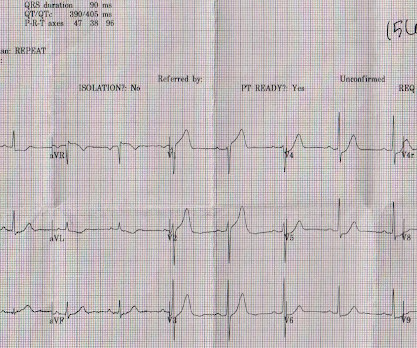
A 50-something man presented in shock with severe chestpain. A 12-lead electrocardiogram, lead V4R , and leads V7-9 were recorded on admission. As discussed earlier — substitution in the chest leads with right-sided and posterior leads confirms RV and posterior MI. The patient was in clinical shock with a lactate of 8.
Submitted by anonymous, written by Pendell Meyers A woman in her 50s presented to the Emergency Department with chestpain and shortness of breath that woke her from sleep, with diaphoresis. See these other cases of arterial pulse tapping artifact: A 60 year old with chestpain Are these Hyperacute T-waves?
Written by Pendell Meyers A middle aged man called EMS for acute chestpain. Physician accuracy in interpreting potential ST-segment elevation myocardial infarction electrocardiograms. EMS recorded this ECG during active symptoms and transmitted it to the ED: I had no information when I was shown the ECG. I said "Not OMI.
This was sent to me by a former resident from a community hospital: A middle-aged woman complained of chestpain and was seen in triage. Comment This paper has received some press recently: Safety of Computer Interpretation of Normal Triage Electrocardiograms The algorithm used was also the GE Marquette 12 SL. Normal ECG."
Case written and submitted by Ryan Barnicle MD, with edits by Pendell Meyers While vacationing on one of the islands off the northeast coast, a healthy 70ish year old male presented to the island health center for an evaluation of chestpain. The chestpain started about one hour prior to arrival while bike riding.
Sent by anonymous A man in his 40s with no previous heart disease presented within 30 minutes of onset of acute chestpain that started while exercising. We have shown many examples of this on this blog. Three patients with chestpain and “normal” ECGs: which had OMI? Chestpain and a computer ‘normal’ ECG.
Written by Jesse McLaren Two 70 year olds had acute chestpain with nausea and shortness of breath, and called paramedics. DIagnostic accuracy oF electrocardiogram for acute coronary OCClusion resulTing in myocardial infarction (DIFOCCULT study). Who needs the cath lab? Int J Cardiol Heart Vasc 2021 2. Aslanger et al.
It is from a 50-something with chestpain: What do you think? That said — followers of Dr. Smith's ECG Blog have already seen numerous clinical cases that we have presented in which the PM Cardio AI Bot app. In a patient with new chestpain — there is no way that the ST-T wave in lead V3 can be normal.
A 70-year-old man calls 911 after experiencing sudden, severe chestpain. Common pitfalls in the interpretation of electrocardiograms from patients with acute coronary syndromes with narrow QRS: a consensus report. This case comes from Sam Ghali ( @EM_RESUS ). Thanks, Sam! This is his 12-Lead ECG: What do you think?
The patient with no prior cardiac history presented in the middle of the night with acute chestpain, and had this ECG recorded during active pain: I did not see any ischemia on this electrocardiogram. This is a case I had quite a while back. See the explainability : She sees large T-waves in V2, V3.
Written by Jesse McLaren A 70 year old with prior MIs and stents to LAD and RCA presented to the emergency department with 2 weeks of increasing exertional chestpain radiating to the left arm, associated with nausea. Echo showed new anterior regional wall motion abnormality and decrease EF from 60% to 45%. Clin Cardiol 2022 4.
Edits by Meyers and Smith A man in his 70s with PMH of hypertension, hyperlipidemia, type 2 diabetes, CVA, dual-chamber Medtronic pacemaker, presented to the ED for evaluation of acute chestpain. Triage ECG: What do you think? This is diagnostic of proximal LAD occlusion. This is a huge anterolateral OMI. I cannot be anything else.
A 61 year-old with chestpain arrived to the ED by ambulance with resolving chestpain. Safety of Computer Interpretation of Normal Triage Electrocardiograms. The chestpain is resolving, so if these are resolving hyperacute T-waves, then followup ECGs should show their size diminishing.
He denied chestpain or shortness of breath. In the clinical context of weakness and fever, without chestpain or shortness of breath, the likelihood of Brugada pattern is obviously much higher. Induced Brugada-type electrocardiogram, a sign for imminent malignant arrhythmias. PM Cardio digitized version.
The utility of the triage electrocardiogram for the detection of ST-segment elevation myocardial infarction. We record ECGs in triage on every patient with chestpain, and some other indications, and this amounts to 8000 ECGs in triage each year, costing at most $200,000 (8000 x $20.00). This paper was just published: Noll S.
can cause ST-segment elevation (STE) on electrocardiogram (ECG), the distinction between them may be hard and complicated. Many researchers, including the editors of this blog, tried to develop such tools in the recent past and we have recommended their use in certain clinical scenarios in many posts on this blog.
An initial electrocardiogram (ECG) is provided below. Although the patient reported experiencing mild pressure-like chestpain, there was suspicion among clinicians that this might be indicative of an older change. See My Comment at the bottom of the page in the May 19, 2020 post in Dr. Smith's Blog ). What do you think?
Cardiology Board Review Question A 48-year-old female with no known medical history presents with acute substernal chestpain. D) An electrocardiogram is most commonly normal in these patients. Patients typically present with acute chestpain, shortness of breath, or syncope. appeared first on BoardVitals Blog.
Because: 1) He has been reading this blog for a long time. Case A 43 year old male with a history of DM II, hyperlipidemia, and a family history of myocardial infarction presented to a family clinic with two days of epigastric pain that started after consuming a meal. 2) He is curious This is how Pendell got started. Normal EKG”.
In the hope of dispelling continued dependence on millimeter-based STEMI criteria — we’ve published numerous cases in recent years in Dr. Smith’s ECG Blog of acute OMI ( O cclusion-based M yocardial I nfarction ) , in which patients have benefited from acute reperfusion despite not satisfying “STEMI criteria”.
This was just published in JAMA Internal Medicine: The de Winter Electrocardiogram Pattern Evolving From Hyperacute T Waves It reminded me that many believe, due to the assertions in the original de Winter's article, that de Winter's waves are stable. He was a 30-something with chestpain. Here is one case of a patient I saw.
This was sent by : Jacob Smith, DO Emergency Medicine Resident Ohio Health Doctors Hospital Emergency Residency Christopher Lloyd, DO, FACEP Director of Clinical Education, USACS Midwest Case A 30 year old patient presents to triage with chestpain. link] Here is the history: A 30 yo man presented complaining of severe chestpain.
A middle aged male with no h/o CAD presented with one week of crescendo exertional angina, and had chestpain at the time of the first ECG: Here is the patient's previous ECG: Here is the patient's presenting ED ECG: There is isolated ST depression in precordial leads, deeper in V2 - V4 than in V5 or V6. There is no ST elevation.
This 42 yo diabetic male presented with cough and foot pain. In spite of aggressive questioning, he denied chestpain, but he did tell one triage nurse that he had had some chest burning, and so he underwent an ECG: There are deep Q-waves and QS-waves in precordial leads V2-V3, with a bit of R-wave left in V4.
The patient contacted EMS after a few hours of chestpain that started 5:30 AM. The pain was described as 6/10 radiating to the right shoulder. The chestpain was described as both sharp and pressure like. Below is the post -PCI electrocardiogram. He is otherwise healthy.
This was a male in his 50's with a history of hypertension and possible diabetes mellitus who presented to the emergency department with a history of squeezing chestpain, lasting 5 minutes at a time, with several episodes over the past couple of months. Plan was for admission for chestpain workup. Patel DJ, et al.
He has never had any chestpain. Explanation: Shown electrocardiogram suggests left ventricular hypertrophy. Shown electrocardiogram suggests left ventricular hypertrophy. The post Hypertropic Cardiomyopathy: A Board Review Question Explained By Video appeared first on BoardVitals Blog. Start with a Free Trial.
She denied chestpain and denied feeling any palpitations, even during her triage ECG: What do you think? 1211-1212 CrossRef View Record in Scopus Google Scholar 2 FI Marcus, W Zareba The electrocardiogram in right ventricular cardiomyopathy/dysplasia. J Electrocardiol, 42 (2009), pp. Figure 3: Key features of idiopathic VT.
She went on to describe her chestpain as a "buffalo sitting on my chest" and a "weird" sensation in her jaw for 1 hour prior to arrival, associated with lightheadedness and diaphoresis. The patient was given fentanyl initially for chestpain with minimal effect and then vomited which was followed by zofran and famotidine.
Smith , d and Muzaffer Değertekin a DIFOCCULT: DIagnostic accuracy oF electrocardiogram for acute coronary OCClUsion resuLTing in myocardial infarction. International Journal of Cardiology Heart & Vasculature Case A 40-year-old man presents with excruciating back pain which has started 1 hour ago.
A middle-aged woman with history of hypertension presented to another hospital approximately 2 hours after onset of chestpain and shortness of breath. Early Continuous ST Segment Monitoring in Unstable Angina: Prognostic Value Additional to the Clinical Characteristics and the Admission Electrocardiogram. mm STE in V1 and 1.5-2.0
The best course is to wait until the anatomy is defined by angio, then if proceeding to PCI, add Cangrelor (an IV P2Y12 inhibitor) I sent the ECG and clinical information of a 90-year old with chestpain to Dr. McLaren. All electrocardiograms (ECGs) and coronary angiograms were blindly analyzed by experienced cardiologists.
He was asked multiple times about chestpain or dyspnea, but repeatedly denied any such symptoms. Patient denied chestpain on initial review of symptoms. Was now endorsing chestpain which began 30 minutes ago. Upon further questioning, he states that he has had intermittent chestpain since yesterday.
A 36 yo male smoker presented to the ED with chestpain. It had started the night before as "indigestion" and had progressed to 8/10 substernal chest pressure radiating to the right shoulder/jaw associated with diaphoresis, nausea, and SOB. This was sent to me by a reader named Aaron. Here is a 15 lead ECG: Sinus rhythm.
male with a history of HTN and ETOH developed squeezing epigastric abdominal pain with associated vomiting and diaphoresis, followed by a syncopal episode which lasted about 10 seconds. When medics arrived, he denied any chestpain, shortness of breath, or palpitations prior to the syncopal episode.
This 60-something with h/o COPD and HFrEF (EF 25%) presented with SOB and chestpain. A deep neural network for 12-lead electrocardiogram interpretation outperforms a conventional algorithm, and its physician over-read, in the diagnosis of atrial fibrillation. The patient in this case presented with dyspnea and chestpain.
A Deep Neural Network learning algorithm outperforms a conventional algorithm for emergency department electrocardiogram interpretation. But lead V2 has a worrisome amount of ST elevation, and in a chestpain patient, I would be worried about STEMI. I do research on Cardiologs' algorithm: Smith SW et al. What an honor.
We organize all of the trending information in your field so you don't have to. Join thousands of users and stay up to date on the latest articles your peers are reading.
You know about us, now we want to get to know you!
Let's personalize your content
Let's get even more personalized
We recognize your account from another site in our network, please click 'Send Email' below to continue with verifying your account and setting a password.


Let's personalize your content