This site uses cookies to improve your experience. To help us insure we adhere to various privacy regulations, please select your country/region of residence. If you do not select a country, we will assume you are from the United States. Select your Cookie Settings or view our Privacy Policy and Terms of Use.
Cookie Settings
Cookies and similar technologies are used on this website for proper function of the website, for tracking performance analytics and for marketing purposes. We and some of our third-party providers may use cookie data for various purposes. Please review the cookie settings below and choose your preference.
Used for the proper function of the website
Used for monitoring website traffic and interactions
Cookie Settings
Cookies and similar technologies are used on this website for proper function of the website, for tracking performance analytics and for marketing purposes. We and some of our third-party providers may use cookie data for various purposes. Please review the cookie settings below and choose your preference.
Strictly Necessary: Used for the proper function of the website
Performance/Analytics: Used for monitoring website traffic and interactions
I see the following: There is sinus tachycardia ( upright P wave with fixed PR interval in lead II ) — at the rapid rate of ~130/minute. See ECG Blog #435 — ECG Blog #313 — as well as My Comment at the bottom of the page in the June 17, 2024 post in Dr. Smith's ECG Blog ). Sinus tachycardia has resolved.
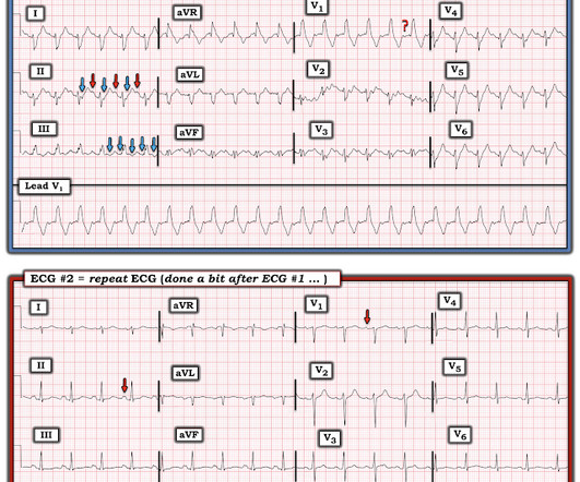
I find AV dissociation in VT to be very difficult to differentiate from artifact, as there are always random blips on tachycardia tracings. Read this post: Idiopathic Ventricular Tachycardias for the EM Physician 2. The 15th beat (2nd beat of V1-V3) appears to be a fusion beat , which is all but diagnostic of VT. Patient intubated.
A previously healthy 53 yo woman was transferred to a receiving hospital in cardiogenicshock. Here was the ECG: There is sinus tachycardia. Referring to Figure-1 — this 53-year old woman who presented in extremis with cardiogenicshock and an initial pH = 6.9, This was sent by a reader. and K was normal.
It shows sinus tachycardia with right bundle branch block. Taking a step back , remember that sinus tachycardia is less commonly seen in OMI (except in cases of impending cardiogenicshock). As per Dr. Frick — sinus tachycardia is usually not seen with acute OMI unless the patient is in cardiogenicshock.
Sinus tachycardia has many potential causes. This is especially true for the elderly patient with sinus tachycardia. What is the cause of the sudden tachycardia? The VSR is what is causing the cardiogenicshock! She had a very elevated troponin T at 12,335 ng/L at the time of presentation.
Figure B At this point, with the ECG changing from diffuse ST depression to widespread ST elevation and the patient presenting in cardiogenicshock, left main coronary artery (LMCA) occlusion is the likely diagnosis. And then, 15 minutes later in today's case — this patient was in cardiogenicshock.
Here are more examples of wide complex tachycardia: these are all a mix of ventricular tachycardia and SVT with aberrancy. He was in cardiogenicshock. There is tachycardia, and there is a wide complex. This wide complex tachycardia could easily be misdiagnosed as V tach.
He was rushed by residents into our critical care room with a diagnosis of STEMI, and they handed me this ECG: There is sinus tachycardia with ST elevation in II, III, and aVF, as well as V4-V6. ACS and STEMI generally do not cause tachycardia unless there is cardiogenicshock. He had this ECG recorded. The HCO3 was 8.
when the usual negative P wave deflection of sinus tachycardia is nowhere to be found in lead V1 )? While of course possible for the rhythm in ECG #1 to be either AFlutter or fascicular VT — sinus tachycardia immediately becomes a much more likely possibility once we know that this patient is critically ill with multisystem disease.
The patient in today’s case presented in cardiogenicshock from proximal LAD occlusion, in conjunction with a subtotally stenosed LMCA. This progressed to electrical storm , with incessant PolyMorphic Ventricular Tachycardia ( PMVT ) and recurrent episodes of Ventricular Fibrillation ( VFib ). RCA — 100% proximal occlussion.
Because of the tachcardia, I would expect her to be very poor left ventricular function and maybe Cardiogenicshock. Still Irregular Blood pressure during these rhythms was adequate; there was no shock. The patient spontaneously converted back to sinus tachycardia. Later, I obtained more clinical history.
There is sinus tachycardia (do not be fooled into thinking this is VT or another wide complex tachycardia!) This pattern is essentially always accompanied by cardiogenicshock and high rates of VT/VF arrest, etc. The patient arrived to the ED in cardiogenicshock but awake. Code STEMI was activated.
We can see enough to make out that the rhythm is sinus tachycardia. Tachycardia is unusual for OMI, unless the patient is in cardiogenicshock (or getting close). As discussed on this blog many times before, proportionality is key to the diagnosis of OMI by ECG. link] I also texted the ECG to Dr. Smith.
Authors' commentary: Cardiogenicshock in the setting of severe aortic stenosis. This patient’s severe aortic stenosis (AS) and associated severe cardiogenicshock likely created the ECG pattern, resulting in a very difficult challenge for our inpatient team. If you can use Doppler, then you can diagnose it.
Among others — See My Comment at the bottom of the page in the September 13, 2024 post of Dr. Smith's ECG Blog ). There is sinus tachycardia at ~100/minute. As often emphasized by Dr. Smith — sinus tachycardia is not a common finding with acute OMI unless something else is going on (ie, cardiogenicshock ).
There is sinus tachycardia. Sinus tachycardia, which exaggerates ST segments and implies that there is another pathology. I have always said that tachycardia should argue against acute MI unless there is cardiogenicshock or 2 simultaneous pathologies. Here is that ECG: What do you think?
The patient died of cardiogenicshock within 24 hours despite mechanical circulatory support. The axis is to the right and QRS complexes in lead I and aVL are predominantly negative suggesting LPFB. This patient at cath had a large CX occlusion with a massive troponin release. Troponin T >42.000ng/L.
The axiom of "type 1 (ACS, plaque rupture) STEMIs are not tachycardic unless they are in cardiogenicshock" is not applicable outside of sinus rhythm. 2) Tachycardia to this degree can cause ST segment changes in several ways. Sometimes you must correct the rhythm to see what lies underneath. Is this inferor STEMI?
The findings include sinus tachycardia, characteristic QRS morphology most diagnostic in V3 with a small R wave followed by a very large S wave with a convex upward ST segment morphology, ST segment strain morphology in the inferior and anterior leads leading to deep symmetric T-wave inversion. and tachycardia, 1.8. incomplete RBBB 1.7
Tachycardia (or nearly) 2. Tachycardia, = 1.8. Finally , they found these independent predictors of PE: Note that tachycardia only has an Odds ratio of 1.8. Tachycardia is unusual in ACS unless there is cardiogenicshock or a second simultaneous pathology. Poor R-wave progression 4. Domed T-wave inversion 5.
Assessment was severe sudden cardiogenicshock. NOTE: For those interested — I review in detail determination of the artifact “culprit extremity” in My Comment in the September 27, 2019 post of Dr. Smith’s ECG Blog. Clinically — the patient was felt to be in cardiogenicshock. What is it? There is STE in V2-V6.
An elderly man with sudden cardiogenicshock, diffuse ST depressions, and STE in aVR Literature 1. We’ve presented many variations on this theme on Dr. Smith’s Blog — with today’s case being distinguished by its discovery on abdominal exam ! A slightly prolonged QTc ( although this is difficult to assess given the tachycardia ).
Why is the patient in shock? He was in profound cardiogenicshock. Both of these features make inferior + RV MI by far the most likely ( Pseudoanteroseptal MI is another name for this ) There is also sinus bradycardia and t he patient is in shock with hypotension. There is an obvious inferior STEMI, but what else?
Here is another proven left main occlusion in a young woman who presented with sudden pulmonary edema, had this ECG recorded, then arrested and was resuscitated after 30 minutes of CPR: This has sinus tachycardia with RBBB and LAFB, and STE in V2-V6 as well as I, aVL This pattern could just as easily be seen in LAD occlusion.
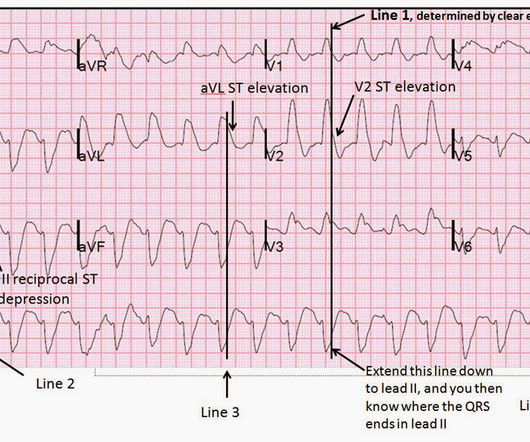
It is a wide complex regular tachycardia at a rate of 120. Is it ventricular tachycardia? I fear that many learners would also not easily recognize where the QRS actually ends, and I fear that some may think that this is ventricular tachycardia due to inability to distinguish QRS from ST segment. The ST Elevation is NOT typical.
The status of the patients chest pain at this time is unknown : EKG 1, 1300: There is sinus tachycardia and artifact of low and high frequency. However, there is also significant tachycardia , with heart rate of 116, and known hypoxia. She arrived to the ED with a nonrebreather mask. Her blood pressure on arrival was 153/69.
We organize all of the trending information in your field so you don't have to. Join thousands of users and stay up to date on the latest articles your peers are reading.
You know about us, now we want to get to know you!
Let's personalize your content
Let's get even more personalized
We recognize your account from another site in our network, please click 'Send Email' below to continue with verifying your account and setting a password.


Let's personalize your content