This site uses cookies to improve your experience. To help us insure we adhere to various privacy regulations, please select your country/region of residence. If you do not select a country, we will assume you are from the United States. Select your Cookie Settings or view our Privacy Policy and Terms of Use.
Cookie Settings
Cookies and similar technologies are used on this website for proper function of the website, for tracking performance analytics and for marketing purposes. We and some of our third-party providers may use cookie data for various purposes. Please review the cookie settings below and choose your preference.
Used for the proper function of the website
Used for monitoring website traffic and interactions
Cookie Settings
Cookies and similar technologies are used on this website for proper function of the website, for tracking performance analytics and for marketing purposes. We and some of our third-party providers may use cookie data for various purposes. Please review the cookie settings below and choose your preference.
Strictly Necessary: Used for the proper function of the website
Performance/Analytics: Used for monitoring website traffic and interactions
As reviewed in ECG Blog #350 — t he clinical significance of Wellens' Syndrome — is that its recognition tells you that the patient has a high-grade LAD narrowing with presumably "hot" thrombus h avin g high propensity to propagate and/or totally occlude the LAD at any point in time ( including immediately ). What is W ellens’ S yndrome ?
The paramedic called the EM physician ahead of arrival and discussed the case and ECGs, and both agreed upon activating "Code STEMI" (even though of course it is not STEMI by definition), so that the acute LAD occlusion could be treated as fast as possible. So the cath lab was activated. Long term outcome is unavailable.
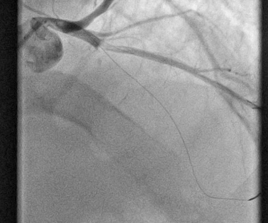
Here is the Queen of Hearts (version 1) interpretation: Click here to sign up for Queen of Hearts Access Angiogram images before and after intervention: Another view (before and after intervention): The patient was confirmed to be in cardiogenicshock, placed on ECMO, also with impella assist device.
A previously healthy 53 yo woman was transferred to a receiving hospital in cardiogenicshock. So Shark Fin really is just a dramatic representation of STEMI, and can be in any coronary distribution. So this is STEMI, right? This was sent by a reader. and K was normal. Here was the ECG: There is sinus tachycardia.
He was rushed by residents into our critical care room with a diagnosis of STEMI, and they handed me this ECG: There is sinus tachycardia with ST elevation in II, III, and aVF, as well as V4-V6. At first glance, it seems the patient is having a STEMI. ACS and STEMI generally do not cause tachycardia unless there is cardiogenicshock.
The VSR is what is causing the cardiogenicshock! Another possible cause of pseudonormalization of T waves mentioned many times on this blog is the pseudonormalization caused by re-occlusion of an infarct related reperfused coronary artery. PIRP is strongly associated with myocardial rupture. Below are two more video files.
The conventional machine algorithm interpreted this ECG as STEMI. Taking a step back , remember that sinus tachycardia is less commonly seen in OMI (except in cases of impending cardiogenicshock). See this post of RV MI with both McConnell sign and "D" sign: Inferior and Posterior STEMI. Both were wrong.
Now appears to be in cardiogenicshock." However, cardiogenicshock usually takes some time to develop, so it is probably subacute." This can only be due to STEMI. Cardiogenicshock and ACS is an indication for the cath lab, even if you don't think there is OMI. I was texted these ECGs.
The ECG shows obvious STEMI(+) OMI due to probable proximal LAD occlusion. The patient in today’s case presented in cardiogenicshock from proximal LAD occlusion, in conjunction with a subtotally stenosed LMCA. The pain was 10/10 in intensity radiating bilaterally to the shoulders and also to the left arm and neck.
Subtle as a STEMI." (i.e., A temporary pacemaker was implanted, and she was admitted to the ICU with cardiogenicshock. In our study, there were 20/53 complete LAD OMI (TIMI-0 flow) which did not meet STEMI criteria. None of the 20 ever evolved to STEMI criteria. This one is easy for the Queen.
Thus, this is BOTH an anterior and inferior STEMI in the setting of RBBB. How old is this antero-inferior STEMI? Although acute anterior STEMI frequently has narrow QR-waves within one hour of onset (1. the presence of such well developed, wide, anterior Q-wave suggests completed transmural STEMI. Could it be acute (vs.
This ECG was read as “No STEMI” with no prior available for comparison. It is true this ECG does not meet STEMI criteria (there is 1.0 The Queen of Hearts sees it of course: Still none of these three ECGs meet STEMI criteria. Do you think we discussed this patient's 2-3 hour delay to reperfusion in our quarterly "STEMI meeting"?
He was in cardiogenicshock. As you will see, this results in the discovery of ST elevation in V2-V4 and I and aVL, diagnostic of anterolateral STEMI. The ED providers activated the cath lab, but the interventionalist refused to come in because it was "not a STEMI". The patient died 8 hours later of cardiogenicshock.
The prehospital and ED computer interpretation was inferior STEMI: There’s normal sinus rhythm, first degree AV block and RBBB, normal axis and normal voltages. The paramedic notes called STEMI into question: “EMS disagree with monitor for STEMI callout. Vitals were normal except for oxygen saturation of 94%. Vitals were normal.
These were catastrophically missed (EM cath lab activation cancelled both times by the interventionalist) Here is a case of Sinus with RBBB and LAFB that was diagnosed correctly by the emergency physicians as acute STEMI. The patient died of cardiogenicshock: Go to the post to see the full explanation.
There is an obvious inferior posterior STEMI(+) OMI. Methods Retrospective study of consecutive inferior STEMI , comparing ECGs of patients with, to those without, RVMI, as determined by angiographic coronary occlusion proximal to the RV marginal branch. What is the atrial activity? Is it sinus arrest with junctional escape?
The ECG was read as "No STEMI" and the patient was treated like an average chest pain patient (despite the fact that a chest pain patient with active pain and active subendocardial ischemia is very high risk). The notes now refer to the patient being in cardiogenicshock, on pressors.
Troponin T peaked at 2074 ng/L (very high, typical of OMI/STEMI). Post PCI the patient became gravely hypotensive and "shocky". She stabilized on dobutamine and levosimendan infusions that could be discontinued after 24 hours. The tricuspid annular plane systolic excursion (TAPSE) improved from 15mm to 19mm within the first 24 hours.
Clinical Course The paramedic activated a “Code STEMI” alert and transported the patient nearly 50 miles to the closest tertiary medical center. 2 The astute paramedic recognized this possibility and announced a CODE STEMI. Authors' commentary: Cardiogenicshock in the setting of severe aortic stenosis. What do you see?
This has been termed a “STEMI equivalent” and included in STEMI guidelines, suggesting this patient should receive dual anti-platelets, heparin and immediate cath lab activation–or thrombolysis in centres where cath lab is not available. aVR ST segment elevation: acute STEMI or not? aVR ST Segment Elevation: Acute STEMI or Not?
My most talented blog readers are paramedics because they have to put themselves on the line every time they activate the cath lab. Then the notes mention "cardiogenicshock" but without any reference to a cardiac echo or to a chest x-ray. Cardiologist note says: "Elevated troponin explained by type II MI due to her shock."
There is an obvious inferior STEMI, but what else? Why is the patient in shock? Besides the obvious inferior STEMI, there is across the precordial leads also, especially in V1. He was in profound cardiogenicshock. This STE is diagnostic of Right Ventricular STEMI (RV MI). A right sided ECG was not recorded.
Assessment was severe sudden cardiogenicshock. In a series of 18 patients with COVID and ST elevation, 8 were diagnosed with STEMI, 6 of whom had an angiogram and it showed obstructive coronary disease. 12 All STEMI patients had very high cTn typical of STEMI (cTnT > 1.0 They recorded an EC G: New ST Elevation.
This pattern is essentially always accompanied by cardiogenicshock and high rates of VT/VF arrest, etc. The patient arrived to the ED in cardiogenicshock but awake. Code STEMI was activated. A man in his 80s with chest pain What, besides large anterior STEMI, is so ominous about this ECG?
When total LM occlusion does present with STE in aVR, there is ALWAYS ST Elevation elsewhere which makes STEMI obvious; in other words, STE is never limited to only aVR but instead it is part of a massive and usually obvious STEMI. All are, however, clearly massive STEMI. This is her ECG: An obvious STEMI, but which artery?
Code STEMI was activated by the ED physician based on the diagnostic ECG for LAD OMI in ventricular paced rhythm. This was several months after the 2022 ACC Guidelines adding modified Sgarbossa criteria as a STEMI equivalent in ventricular paced rhythm). LAFB, atrial flutter, anterolateral STEMI(+) OMI. Limkakeng AT.
When the ST vector is primarily posterior, the diagnosis is usually posterior STEMI. ST depression maximal in V5 and V6 cannot be reciprocal to subepicardial, transmural ischemia under aVR because, as stated above, there is no ventricular myocardium beneath that lead, no STEMI under aVR. I just read Ken's comments before publishing.
The patient was brought to the ED as a possible Code STEMI and was seen directly by cardiology. Similarly, STEMI guidelines call for urgent angiography for refractory ischemia or electrical/hemodynamic instability, regardless of ECG findings. But by this time the patient went into cardiogenicshock and passed away.
A recent study found that SCAD causes almost 20% of STEMI in young women. examined SCAD presenting as STEMI (unlike Hassan et al. were pretty sick, with mostly LM/pLAD lesions and high rates of cardiogenicshock. I had no idea SCAD was so common a cause of acute STEMI in younger women, even when they are non-smokers.
He was in cardiogenicshock requiring an impella for several days after cath. We've shown many cases on Dr. Smith's ECG Blog of subtle ECG findings that rapidly evolve into dramatic ST-T wave changes. No further troponins were measured. But we should not be surprised.
Because of the tachcardia, I would expect her to be very poor left ventricular function and maybe Cardiogenicshock. Dyspnea, Right Bundle Branch block, and ST elevation Here are two more cases where the differential diagnosis is acute OMI vs. LV aneurysm: Is this acute STEMI? Not all anterior LV aneurysm has a QS-wave.
I have always said that tachycardia should argue against acute MI unless there is cardiogenicshock or 2 simultaneous pathologies. Exclusion criteria were age less than 18, SBP less than 100 mmHg, echocardiogram with EF less than 50%, STEMI, pregnancy, and trauma. PR depression, which suggests pericarditis 4.
The axiom of "type 1 (ACS, plaque rupture) STEMIs are not tachycardic unless they are in cardiogenicshock" is not applicable outside of sinus rhythm. Is that an obvious STEMI underneath that rhythm? Is this inferor STEMI? Atrial Flutter with Inferior STEMI? If I fix the rhythm will the ST changes resolve?
PCI mid LCx So this is an OMI (Occlusion Myocardial Infarction), but not a STEMI Echo: Decreased left ventricular systolic performance, mild/moderate. The patient went into cardiogenicshock and ultimately died of this MI. Angiogram: LM 30% ostial. LAD 80% mid LCx occluded mid (acute infarct lesion) RCA 80% mid. Sandoval Y.
See our other cases with similar patters, to burn this deep into your brain files: Smith : In my experience, these cases of LAD OMI with RBBB and LAFB are either about to arrest, post-arrest, and/or in cardiogenicshock. Acute chest pain, right bundle branch block, no STEMI criteria, and negative initial troponin. To quote Drs.
Whenever there is tachycardia, I am skeptical of OMI unless it has led to severely compromised ejection fracction with cardiogenicshock. Supply-demand mismatch can cause ST Elevation (Type 2 STEMI). Also see these posts of Type II STEMI. Truly, the Marquette 12 SL algorithm correctly identifies this STEMI.
We organize all of the trending information in your field so you don't have to. Join thousands of users and stay up to date on the latest articles your peers are reading.
You know about us, now we want to get to know you!
Let's personalize your content
Let's get even more personalized
We recognize your account from another site in our network, please click 'Send Email' below to continue with verifying your account and setting a password.


Let's personalize your content