This site uses cookies to improve your experience. To help us insure we adhere to various privacy regulations, please select your country/region of residence. If you do not select a country, we will assume you are from the United States. Select your Cookie Settings or view our Privacy Policy and Terms of Use.
Cookie Settings
Cookies and similar technologies are used on this website for proper function of the website, for tracking performance analytics and for marketing purposes. We and some of our third-party providers may use cookie data for various purposes. Please review the cookie settings below and choose your preference.
Used for the proper function of the website
Used for monitoring website traffic and interactions
Cookie Settings
Cookies and similar technologies are used on this website for proper function of the website, for tracking performance analytics and for marketing purposes. We and some of our third-party providers may use cookie data for various purposes. Please review the cookie settings below and choose your preference.
Strictly Necessary: Used for the proper function of the website
Performance/Analytics: Used for monitoring website traffic and interactions
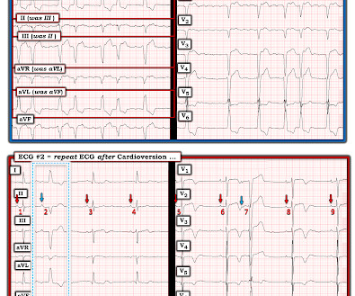
By the P s, Q s, 3 R Approach ( See ECG Blog #185 ): The rhythm is fast and QRS complexes are R egular. PEARL # 4: As emphasized in ECG Blog #204 , in which I review derivation of the bundle branch blocks — RBBB is a terminal conduction delay. Figure-2: I've labeled the initial ECG in today's case. And a Final Tracing.
Discontinue all negative chronotropic agents, since the risk of torsade is much higher with bradycardia or pauses. Smith Pacing in today's case is an effective intervention as doing so prevents the bradycardia and pauses that are likely to precipitate additional episodes of Torsades de Pointes. ( The plan: 1. J Am Coll Cardiol.
It's important to appreciate that we can not rule out the possibility that this rhythm represents Mobitz II 2nd-degree AV block, in which case the patient will need a pacemaker ( See ECG Blog #236 — for review on the ECG diagnosis of the 2nd-degree AV Blocks ). ECG Blog #192 — The 3 Causes of AV Dissociation. STEP-by-STEP. .
to 1828 msec. ) — which corresponds to a variation in the rate of sinus bradycardia from 36-to-33/minute. This makes sense given that the underlying rhythm in today's case appears to be marked sinus bradycardia and arrhythmia , with a ventricular escape rhythm appearing when the SA node rate drops below 33/minute.
There was concern that the rhythm might represent ventricular tachycardia, so lidocaine was given and one attempt at cardioversion was performed. See our other countless hyperkalemia cases below: General hyperkalemia cases: A 50s year old man with lightheadedness and bradycardia Patient with Dyspnea. A Very Wide Complex Tachycardia.
Shortly after isoprenalin infusion was initiated, there were short runs of ventricular tachycardia. For example — bradycardia and AV conduction disturbances are not uncommon with Hyperkalemia , with these conduction disturbances most often resolving once serum K+ is corrected. She was started on isoprenalin (isoproterenol).
EMS reports intermittent sinus tachycardia and bradycardia secondary to some type of heart block during transport. Smith comment: Go here for a comprehensive blog post on syncope and link to the most detailed version of the Canadian Syncope Rule: Emergency Department Syncope Workup. See these blog posts.
This progressed to electrical storm , with incessant PolyMorphic Ventricular Tachycardia ( PMVT ) and recurrent episodes of Ventricular Fibrillation ( VFib ). There was no evidence bradycardia leading up to the runs of PMVT ( as tends to occur with Torsades ). He required multiple defibrillations within a period of a few hours.
The ECG shows sinus tachycardia with RBBB and LAFB, without clear additional superimposed signs of ischemia. The Initial ECG in Today's Case: As per Dr. Meyers — the initial ECG in today's case shows sinus tachycardia with bifascicular block ( = RBBB/LAHB ). Sinus Tachycardia ( common in any trauma patient. ).
During the night, while on telemetry, the patient became bradycardic, with periods of isorhythmic AV dissociation (nodal escape rhythm alternating with sinus bradycardia), and there were sporadic PVCs. The above ECGs show the initiation and continuation of a polymorphic ventricular tachycardia.
This blog explores the ways wearable technology can help track heart health, the advantages it offers, and how it contributes to better outcomes for those requiring surgical intervention. Early detection of conditions like AFib, bradycardia, or tachycardia allows patients to address issues before they become critical.
Both of these features make inferior + RV MI by far the most likely ( Pseudoanteroseptal MI is another name for this ) There is also sinus bradycardia and t he patient is in shock with hypotension. A narrow complex bradycardia without any P-waves is also likely to respond to atropine, as it may be a junctional rhythm.
Wide-complex tachycardia: VT or aberrant, or "other?" A wide-complex tachycardia in an older patient must immediately suggest ventricular tachycardia. Sinus tachycardia with aberrancy was unlikely as the rate was consistently 150 bpm, without spontaneous variation. second ), in which the tachycardia is sustained.
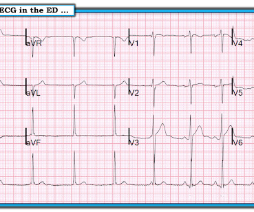
Initial ECG in the ED: Presenting ECG : Wide-complex tachycardia at a rate about 200. This is overwhelmingly likely to be ventricular tachycardia, even if only age and medical history are considered. Nevertheless, the widths of both the QRS complex and the RS duration are similar in both the old ECG and the tachycardia.
A 50-something male with unspecified history of cardiomyopathy presented in diabetic ketoacidosis (without significant hyperkalemia) with a wide complex tachycardia and hypotension. Analysis: there is a wide complex tachycardia. This was the interpretation I put into the system: WIDE COMPLEX TACHYCARDIA. It is regular.
ECG is consistent with severe hypokalemia and/or hypomagnesemia causing prolonged QT (QU) at high risk of Torsades (which is polymorphic ventricular tachycardia in the setting of a long QT interval). Polymorphic Ventricular Tachycardia Long QT Syndrome with Continuously Recurrent Polymorphic VT: Management Cardiac Arrest. Is it STEMI?
Here was his initial ED ECG: There is sinus tachycardia at a rate of about 140 There is profound ST Elevation across all precordial leads, as well as I and aVL. I said I think there is a fixed stenosis in the LAD and the tachycardia and stress caused a type 2 STEMI.
My Comment , by K EN G RAUER, MD ( 7/5/2018 ): This blog post provides an excellent example of how a patient with SSS ( = S ick S inus S yndrome ) may present. during which sinus bradycardia and arrhythmia are seen but not to a degree that produces symptoms. The indication for pacemaker placement with SSS is symptomatic bradycardia.
We've presented numerous examples of hyperkalemia on Dr. Smith's ECG Blog. Descriptive analysis of the ECG in today's case reveals a regular, narrow tachycardia at ~130/minute , without clear sign of sinus P waves. But the rate is ~130/minute — which is a bit fast for sinus tachycardia. WHY Isn't the QRS Wide?
As per Dr. Nossen — today's initial ECG ( LEFT tracing in Figure-2 ) shows sinus bradycardia with QRS widening due to bifascicular block ( RBBB/LAHB ). There is marked, diffuse ST segment depression in ECG #1.
This has been discussed many times before on this blog. In-depth discussion is beyond the scope of this blog. Automatic activity refers to enhanced pacemaking function (typically from a non sinus node source), for example atrial tachycardia. The rhythm is AIVR -- accelerated idioventricular rhythm. Do not treat AIVR.
Other than tachycardia, Other than slight tachycardia, vitals were within normal limits (including oxygen saturation). Like other cases on this blog who died before cath was done, I cannot prove that this patient had OMI. Sent by anonymous, written by Pendell Meyers A middle aged man presented with acute shortness of breath.
He had multiple episodes of bradycardia and nonsustained ventricular tachycardia. Serial high sensitivity troponin T (URL 15 ng/L) values were negative and stagnant. Patient 1 remained in the hospital overnight. He went to the cath lab at 0900 the next morning. It was stented.
After initiating treatment for hyperkalemia, repeat ECG showed resolution of Brugada pattern: The ECG shows sinus tachycardia. ST elevation in aVL with reciprocal ST depression in the inferior leads Shock, bradycardia, ST Elevation in V1 and V2. He also received insulin with D50, sodium bicarbonate, and kayexalate for hyperkalemia.
A rapid heartbeat (tachycardia) can increase the workload on your heart, while a slow heartbeat (bradycardia) can cause fatigue and dizziness. It’s crucial to monitor your blood pressure regularly and report any significant changes to your healthcare provider so they can adjust the dosage of the medication accordingly.
I completely agree with Dr. Nossen that in this patient with new CP and sinus tachycardia with LAHB — that the T waves in each of the inferior leads are hyperacute ( ie, clearly disproportionately "bulky" given size of the QRS in these leads ). — and which other patient(s) need to be seen as soon as possible to figure out what is going on?
This phenomenon is best known as, A V D issociation b y D efault ie, default of the sinus pacemaker due to marked sinus bradycardia, which transiently allows the AV nodal escape pacemaker to take over. Instead, as we state above the rhythm is sinus bradycardia with marked sinus arrhythmia. Close follow-up is essential.
I have periodically called attention to examples of the Ashman phenomenon as they occur in Dr. Smith's ECG Blog ( See My Comments in the January 5, 2020 post — the June 17, 2020 post — and the March 30, 2023 post , among others ). But there are 3 other wide beats in the tachycardia that begins with beat #6 ( = beats #7; 13,14 ).
There is also bradycardia. Bradycardia puts patients at risk for "pause-dependent" Torsades de Pointes. Torsades in acquired long QT is much more likely in bradycardia because the QT interval following a long pause is longer still. See here for management of Polymorphic Ventricular Tachycardia , which includes Torsades.
Prior ECG on file: Sinus tachycardia, imperfect baseline, otherwise unremarkable. Sinus tachycardia with unequivocal evidence of hyperkalemia, including widened QRS with "pulled apart" morphology (widened QRS) compared to baseline, as well as clearly peaked T-waves.
As we've discussed on numerous other posts in Dr. Smith's ECG Blog ( See My Comment at the bottom of the page in the May 5, 2022 post) — a growing number of conditions other than Brugada Syndrome have been found to temporarily produce a Brugada-1 ECG pattern. Sinus Tachycardia ( common in any trauma patient. ).
Otherwise vitals after intubation were only notable for tachycardia. An initial EKG was obtained: Computer read: sinus tachycardia, early acute anterior infarct. She was ventilated by bag-valve-mask by EMS on arrival and was quickly intubated with etomidate and succinylcholine. A rectal temperature was obtained which read 107.9
A useful classification of WCT ( W ide- C omplex T achycardia ) rhythms — separates them into those that are mono morphic ( with similar QRS morphology during the tachycardia ) vs those that are poly morphic ( in which QRS morphology varies ). QUESTION Regarding ECG #5: WHAT ECG findings in ECG #5 predispose to development of Torsades?
U waves may also be found in patients with LVH and/or bradycardia , or occasionally as a normal variant. Y OU s hould h ave n oted the following additional ECG findings in ECG #1 : There is sinus tachycardia at ~100/minute in ECG #1. N OTE # 1 — U waves are not specific for hypokalemia! second ).
My L IST includes the following: i ) LVH with strain; ii ) Ischemia; iii ) Digoxin use; iv ) HypoKalemia and/or HypoMagnesemia; v ) Tachycardia; and , vi ) Any combination of i-thru-v. In my experience, Ive seen U waves not only with low K+/low Mg++ but also in patients with bradycardia, LVH, and sometimes in normal subjects.
There is a regular, wide complex, (mostly) monomorphic tachycardia. The differential of wide, regular, monomorphic tachycardia is: VT or SVT with aberrancy, all +/- hyperkalemia (see diagrams below). Really wide tachycardia = VT or SVT with conduction abnormality + hyperkalemia, until proven otherwise.
There is sinus tachycardia and also a large R-wave in aVR. Drug toxicity , especially diphenhydramine , which has sodium channel blocking effects, and also anticholinergic effects which may result in sinus tachycardia, hyperthermia, delirium, and dry skin. Her temperature was 106 degrees. As part of the workup, she underwent an ECG.
This ECG shows a sinus bradycardia with a normal conduction pattern (normal PR, normal QRS, and normal QTc), normal axis, normal R-wave progression, normal voltages. Hypothermia can also produce bradycardia and J waves, with a pseudo-STEMI pattern. This is the 2nd ECG from the February 8, 2022 post in Dr. Smith's ECG Blog ).
In view of the clinical history ( ie, that this patient had a history of longstanding AFlutter but as far as we know, no chest pain ) this ST-T wave change most likely reflects a "memory" effect , in which there will often be ST-T wave abnormalities that persist for hours ( up to a few days ) following a long period of a sustained tachycardia.
MY Thoughts on ECG #1: The rhythm is sinus bradycardia at a rate just under 60/minute. I've reviewed on many occasions in this ECG Blog — cases in which acute OMIs were overlooked and misdiagnosed as NSTEMI ( See ECG Blog #400 — among many others ). There is sinus tachycardia at ~100/minute — with diffuse ST-T wave depression.
If the patient has Abnormal Vital Signs (fever, hypotension, tachycardia, or tachypnea, or hypoxemia), then these are the primary issue to address, as there is ongoing pathology which must be identified. Thus, if there is documented sinus bradycardia, and no suspicion of high grade AV block, at the time of the syncope, this is very useful.
During observation in the ED the patient had multiple self-terminating runs of Non-Sustained monomorphic Ventricular Tachycardia (NSVT). The intricacies of the different classes of antiarrhythmics and their mechanism of action extend beyond the scope of this blog. Potassium and magnesium serum levels were normal. No PVCs are seen.
There was never ventricular fibrillation (VF) or ventricular tachycardia (VT), no shockable rhythm. Here is a similar case: Collapse, Ventricular Tachycardia, Cardioverted, Comatose on Arrival. Agitation, Confusion, and Unusual Wide Complex Tachycardia. There is sinus tachycardia at ~115/minute.
Within ten minutes, she developed bradycardia, hypotension, and ST changes on monitor. Bradycardia and heart block are very common in RCA OMI. There was indication of parasympathetic overdrive ( the acute inferior STEMI with profound bradycardia and junctional escape ). He told the patient this horrible news.
We organize all of the trending information in your field so you don't have to. Join thousands of users and stay up to date on the latest articles your peers are reading.
You know about us, now we want to get to know you!
Let's personalize your content
Let's get even more personalized
We recognize your account from another site in our network, please click 'Send Email' below to continue with verifying your account and setting a password.


Let's personalize your content