This site uses cookies to improve your experience. To help us insure we adhere to various privacy regulations, please select your country/region of residence. If you do not select a country, we will assume you are from the United States. Select your Cookie Settings or view our Privacy Policy and Terms of Use.
Cookie Settings
Cookies and similar technologies are used on this website for proper function of the website, for tracking performance analytics and for marketing purposes. We and some of our third-party providers may use cookie data for various purposes. Please review the cookie settings below and choose your preference.
Used for the proper function of the website
Used for monitoring website traffic and interactions
Cookie Settings
Cookies and similar technologies are used on this website for proper function of the website, for tracking performance analytics and for marketing purposes. We and some of our third-party providers may use cookie data for various purposes. Please review the cookie settings below and choose your preference.
Strictly Necessary: Used for the proper function of the website
Performance/Analytics: Used for monitoring website traffic and interactions
To do this — I apply the P s, Q s, 3 R Approach ( See ECG Blog #185 — for review of my system ). Even if we stopped here — We could conclude the following: There is marked bradycardia in today's rhythm ( ie, Heart rate in the low 30s ). Figure-4: This is the laddergram I favor for illustrating the mechanism of today's rhythm.
I see the following: The rhythm is sinus bradycardia at ~55-60/minute. PEARL # 1: As I've emphasized often in this ECG Blog — the course of acute MI from acute coronary occlusion — is often staggered. PEARL # 1: As I've emphasized often in this ECG Blog — the course of acute MI from acute coronary occlusion — is often staggered.
That said — obvious findings include: i ) Marked bradycardia! — This suggests ischemia of uncertain duration. Given this patient's older age — if nothing "fixable" is found, she most likely has SSS ( S ick S inus S yndrome ) and will need a pacemaker ( See ECG Blog #342 for more on SSS ). be regular! —
Discontinue all negative chronotropic agents, since the risk of torsade is much higher with bradycardia or pauses. It should be kept in mind that on occasions, beta-one agonist can result in increased ventricular ectopy e.g., in severe myocardial ischemia (by increasing myocardial demand), or sometimes with congenital long-QT syndrome.
to 1828 msec. ) — which corresponds to a variation in the rate of sinus bradycardia from 36-to-33/minute. This makes sense given that the underlying rhythm in today's case appears to be marked sinus bradycardia and arrhythmia , with a ventricular escape rhythm appearing when the SA node rate drops below 33/minute.
The ECG does not show any definite signs of ischemia. It is unclear if the patient was pain free at this time. In fact, the ECG was described as normal, and without serial ECGs or prior ECGs for comparison it could be. Initial high sensitivity troponin I returned at 6ng/L (normal 0.20
See our other blog posts of hypothermia and Osborn waves -- Massive Osborn Waves of Severe Hypothermia (23.6 Altered Mental Status, Bradycardia == MY Comment , by K EN G RAUER, MD ( 2/2 /2024 ): == Dr. Meyers began today’s case with the clinical challenge of asking you to identify the underlying cause of ECG #2. Is there a long QT?
No ischemia. Case continued Another ECG was recorded 3 hours later, still 1/10 pain: There is sinus bradycardia with RBBB. This is a conundrum, because it is clear that the patient is having an acute MI, the ECG is dynamic, but the pain is very mild and there is no ECG evidence of active transmural ischemia.
There is no definite evidence of acute ischemia. (ie, Simply stated — t he patient was having recurrent PMVT without Q Tc prolongation, and without evidence of ongoing transmural ischemia. ( Some residual ischemia in the infarct border might still be present. Both episodes are initiated by an "R-on-T" phenomenon.
Common explanations for unusual rhythms such as this one include: i ) Hyperkalemia ( or other severe electrolyte disorder ); ii ) Recent infarction/ischemia; iii ) Sleep apnea; iv ) Severe hypothyroidism; v ) Acute neurologic catastrophe (ie, stroke, bleed, trauma, tumor ); vi ) Some other toxicity.
Her vital signs were within normal limits except for bradycardia at 55 bpm. It is probably sinus bradycardia with very small/depressed P-waves and prolonged PR interval. P EARL # 4 In my opinion, it is not worth wasting time trying to figure out the specific rhythm diagnosis of a bradycardia when there is hyperkalemia.
I will leave more detailed rhythm discussion to the illustrious Dr. Ken Grauer below, but this use of calipers shows that the rhythm interpretation is: Sinus bradycardia with a competing (most likely junctional) rhythm. The fact that R waves 2 through 6 are junctional does make ischemia more difficult to interpret -- but not impossible.
For instance, if there were inappropriate sinus bradycardia at less than 60 bpm, the atrial pacer would take over if it is programmed to wait 1 second before firing. The T-waves of both of these beats have, coincidentally , a superimposed P-wave Clinical course: The potassium was normal, there was no ischemia or drug toxicity.
The patient with no prior cardiac history presented in the middle of the night with acute chest pain, and had this ECG recorded during active pain: I did not see any ischemia on this electrocardiogram. The patient has active chest pain, so if these are abnormally large T-waves This link shows 13 blog posts of Posterior Reperfusion T-waves.
A prior ECG was available for comparison: Normal One might be tempted to interpret the ST depression as ischemia, but as Smith says, "when the QT is impossibly long, think of hypokalemia and a U-wave rather than T-wave." QUESTION #2: If it were not for the markedly prolonged QTc — Wouldn't ECG #1 look like diffuse subendocardial ischemia?
Both of these features make inferior + RV MI by far the most likely ( Pseudoanteroseptal MI is another name for this ) There is also sinus bradycardia and t he patient is in shock with hypotension. A narrow complex bradycardia without any P-waves is also likely to respond to atropine, as it may be a junctional rhythm.
Below is the first ECG recorded by paramedics after 2 hours of chest pain, interpreted by the machine as “possible inferior ischemia”. There’s competing sinus bradycardia and junctional rhythm, with otherwise normal conduction, borderline right axis, normal R wave progression and voltages. What do you think?
The first task when assessing a wide complex QRS for ischemia is to identify the end of the QRS. The ST segment changes are compatible with severe subendocardial ischemia which can be caused by type I MI from ACS or potentially from type II MI (non-obstructive coronary artery disease with supply/demand mismatch). What do you think?
During the night, while on telemetry, the patient became bradycardic, with periods of isorhythmic AV dissociation (nodal escape rhythm alternating with sinus bradycardia), and there were sporadic PVCs. Most such rhythms in the setting of ischemia are VF and will not convert without defibrillation. Acute ischemia?
The STD maximal in V1-V4 is diagnostic of acute transmural posterior wall ischemia, most likely due to posterior OMI. Subendocardial ischemia does not localize, and subendocardial ischemia presents with STD maximal in V5-6, II, and STE in aVR. Subendocardial ischemia does not localize.
The ECG shows sinus tachycardia with RBBB and LAFB, without clear additional superimposed signs of ischemia. I've copied KEY points from My Comment in the August 6, 2022 post in Dr. Smith's ECG Blog — regarding the answer to this question. Chest trauma was suspected on initial exam. Figure-1: The initial ECG in today's case.
Due to bradycardia, a 12-lead ECG was obtained: There is atrial fibrillation at a rate of 54. But because of bradycardia, a 12-lead was obtained, which gave the critical diagnosis. Slow atrial fibrillation implies an sick AV node, or one affected by electrolytes, ischemia, or medications/drugs. His breath alcohol was 0.259.
Remember, in diffuse subendocardial ischemia with widespread ST-depression there may b e ST-E in lead s aVR and V1. There are well formed R-waves with good voltage/amplitude which is uncommon for ischemia. The ECG does not show any signs of ischemia. True Positive ECG#2 : Also sinus rhythm. There is ST depression in V1.
My Comment , by K EN G RAUER, MD ( 7/5/2018 ): This blog post provides an excellent example of how a patient with SSS ( = S ick S inus S yndrome ) may present. during which sinus bradycardia and arrhythmia are seen but not to a degree that produces symptoms. The indication for pacemaker placement with SSS is symptomatic bradycardia.
Description Sinus bradycardia. There is ST elevation in V2 and V3 There are inverted T-waves in V2 and V3 There are prominent U-waves in V2 and V3 Many responders were worried about ischemia or hypertrophic cardiomyopathy. This short QT at least makes ischemia all but impossible. There is high voltage. This is a normal variant.
If you put the inferior and posterior findings together, it is diagnostic of OMI This was read as "inferior ischemia" without any other information by Dr. Richard Gray and as probable reperfused inferior-posterior OMI much later by both me and Pendell Meyers, also without any clinical information.
This has been discussed many times before on this blog. In-depth discussion is beyond the scope of this blog. Possible mechanisms of ventricular arrhythmias elicited by ischemia followed by reperfusion. The rhythm is AIVR -- accelerated idioventricular rhythm. Do not treat AIVR. In fact, use of antidyrhythimcs (e.g., Moffat, M.
Sinus bradycardia, normal conduction, normal axis, normal R wave progression, no hypertrophy. 2] Here there is no posterior ST elevation, but the anterior ST depression is also less—so it is dynamic, confirming acute ischemia. What do you think? But it is still STEMI negative.
After the heart rate increased slightly, here was the repeat ECG: Sinus bradycardia, only slightly faster rate than prior. Learning Points: Ectopic atrial rhythm can produce atrial repolarization findings that can be confused for acute ischemia, STEMI, or OMI.
Extensive conduction system abnormalities can have various causes (ischemia, genetic, infectious, amyloid, etc). For example — bradycardia and AV conduction disturbances are not uncommon with Hyperkalemia , with these conduction disturbances most often resolving once serum K+ is corrected.
Soon afterward, the patient’s symptoms return along with lightheadedness, bradycardia, and hypotension. The patient has also developed sinus bradycardia, which may result from right coronary artery ischemia to the SA node. The Queen of Hearts agrees: Around this time his initial high sensitivity troponin I resulted at 231 ng/L.
Whether these EKGs show myocarditis, a normal variant, or something else, they are overall not typical of transmural ischemia of the anterior or high lateral walls. He had multiple episodes of bradycardia and nonsustained ventricular tachycardia. The patient had none of these conditions. Patient 1 remained in the hospital overnight.
Osborn waves have been reported with hypercalcemia, brain injury, subarachnoid hemorrhage, Brugada syndrome, cardiac arrest from VFib — and — severe, acute ischemia resulting in acute MI ( See My Comment in the November 22, 2019 post on Dr. Smith’s Blog ). Rituparna et al — as well as Chauhan and Brahma ( Int.
There are 3 etiologies I always think of with bradycardia and AV block: 1. There was no evidence of ischemia. In addition to ruling out rate-slowing medication serum electrolyte disorders and/or ischemia/infarction as potential causes of bradyarrhythmias one should also rule out hypothyroidism + sleep apnea. Hyperkalemia.
Here is his previous ECG: This was my interpretation of the first ECG: Sinus bradycardia with less than 1mm ST elevation in V4-V6, elevated compared to the previous ECG, suggestive of lateral MI. This is his first ECG in the department, which I saw as it was being printed: What do you think? Notice how useful serial ECGs are!
My most talented blog readers are paramedics because they have to put themselves on the line every time they activate the cath lab. There is a junctional bradycardia. Furthermore, there are T-wave changes in V2 and V3 which are highly suggestive of ischemia, but difficult to localize: anterior? And they teach me a lot.
Hyperkalemia causes peaked T waves and the "killer B's of hyperkalemia", including bradycardia, broad QRS complexes, blocks of the AV node and bundle branches, Brugada morphology, and otherwise bizarre morphology including sine wave. With a twist. Do you recognize this ECG yet? Right Bundle Branch Block with ST Elevation in V1?
Followup ECG: No Change Absence of evolution is the best evidence against ischemia as the etiology. I was taught that the tell-tale sign of ischemia vs an electrical abnormality was in the hx, i.e. chest pain for the ischemia and potential syncope for brugada. Ischemia/infarction. Bradycardia. Hypothermia.
2] Conduction through the accessory pathway can be intermittent (with different degrees of pre-excitation), and affected by ischemia. 3] So a patient with WPW can have the pattern induced by ischemia, and there is also a report of a patient with pre-existing WPW which was “ablated” by myocardial infarction after an LAD occlusion.[4]
Diffuse ST depression with ST elevation in aVR: Is this pattern specific for global ischemia due to left main coronary artery disease? Ischemia b. ST depression: is it ischemia? In my experience, Ive seen U waves not only with low K+/low Mg++ but also in patients with bradycardia, LVH, and sometimes in normal subjects.
In any case, there is bradycardia. There is ST depression beyond the end of the wide QRS in I, II, aVF, and V4-V6, diagnostic of with subendocardial ischemia. due to PE) may result in STEMI (and, if anterior, it can be from anterior LV or anterior RV ischemia, or both) from low coronary pressure and flow, simply due to the shock.
Triage physician interpretation: -sinus bradycardia -lateral ST depressions While there are lateral ST depressions (V5, V6) the deepest ST depressions are in V4. Ischemic ST-Segment Depression Maximal in V1-V4 (Versus V5-V6) of Any Amplitude Is Specific for Occlusion Myocardial Infarction (Versus Nonocclusive Ischemia). 121.022866.
Here are inferior leads, and aVL, magnified: A closer inspection of the inferior leads and aVL Sinus bradycardia. I had no history on the case and no prior ECG for comparison. What do you think? The T-wave in lead III is slightly tall and broad (increased area under the curve) compared to its QRS complex.
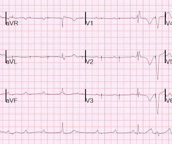
Here is his ECG: There is no clear evidence of OMI or ischemia. The Initial ECG in Today's Case: ECG #1 showed sinus bradycardia at a rate slightly under 60/minute — normal intervals — slight left axis ( about -15 degrees ) — and no chamber enlargement. A 40-something male with no previous cardiac disease presented with chest pain.
We organize all of the trending information in your field so you don't have to. Join thousands of users and stay up to date on the latest articles your peers are reading.
You know about us, now we want to get to know you!
Let's personalize your content
Let's get even more personalized
We recognize your account from another site in our network, please click 'Send Email' below to continue with verifying your account and setting a password.


Let's personalize your content