This site uses cookies to improve your experience. To help us insure we adhere to various privacy regulations, please select your country/region of residence. If you do not select a country, we will assume you are from the United States. Select your Cookie Settings or view our Privacy Policy and Terms of Use.
Cookie Settings
Cookies and similar technologies are used on this website for proper function of the website, for tracking performance analytics and for marketing purposes. We and some of our third-party providers may use cookie data for various purposes. Please review the cookie settings below and choose your preference.
Used for the proper function of the website
Used for monitoring website traffic and interactions
Cookie Settings
Cookies and similar technologies are used on this website for proper function of the website, for tracking performance analytics and for marketing purposes. We and some of our third-party providers may use cookie data for various purposes. Please review the cookie settings below and choose your preference.
Strictly Necessary: Used for the proper function of the website
Performance/Analytics: Used for monitoring website traffic and interactions
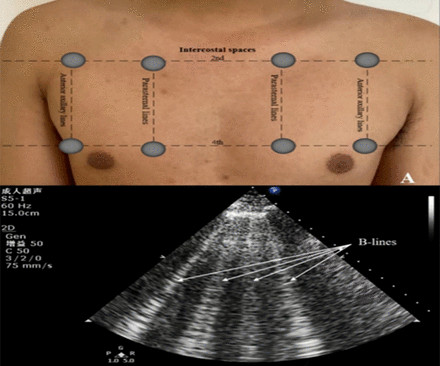
Objectives Prognostic impact of lung ultrasound-derived B-lines (LUS-BL) in heart failure with mildly reduced left ventricular ejection fraction (HFmrEF) patients remains elusive. to 2.94), increased ratio of early transmitral flow velocity to early mitral annular velocity (>24, HR=1.79, 95% CI 1.11
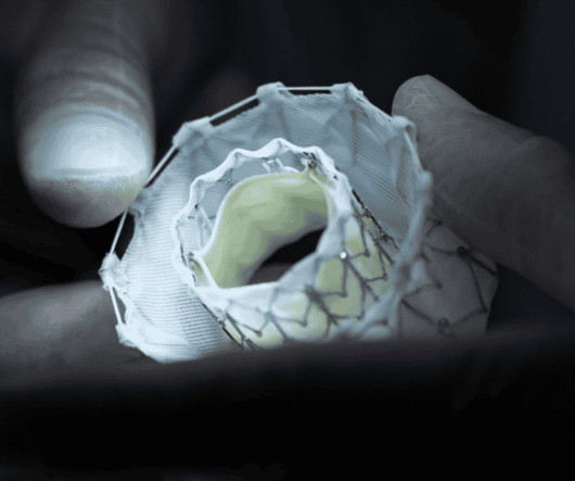
Goar is internationally recognized for remarkable career achievements which include the development of the MitraClip , a medical innovation used worldwide to reduce mitral regurgitation, a common heart condition that occurs when the mitral valve doesn't close properly.
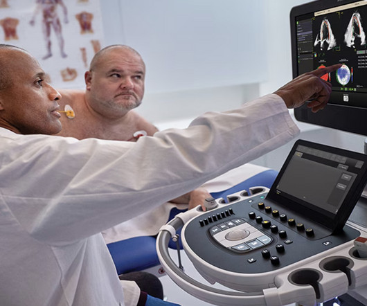
Philips’ ultrasound AI strategy took another big step this week, with the launch of its next-generation echo AI platform, which will come integrated with the company’s cardiovascular ultrasound systems and bring a range of new echo-automating capabilities.
Food and Drug Administration (FDA) has granted 510(k) clearance for its first-of-a-kind, AI-powered AISAP CARDIO point-of-care ultrasound (POCUS) software platform. We know that structural heart disease and heart failure are the leading causes of hospitalization and morbidity in the U.S.
Introduction:Fabry disease, caused by mutations in the gene encoding alpha-galactosidase A, results in the accumulation of globotriaosylceramide (Gb3), leading to systemic complications including cardiac involvement such as mitral regurgitation (MR).The
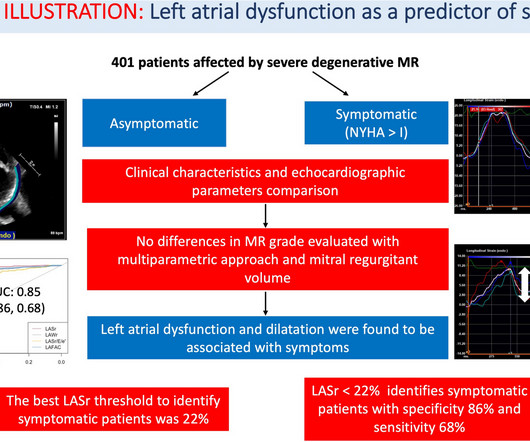
In patients affected by mitral regurgitation (MR) an impairment in LA compliance and. Left atrium (LA) is far from simply being a passive connection chamber between left ventricle and the pulmonary circulation.
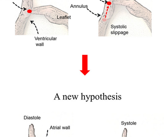
Timely electrical activation of the left ventricular (LV) papillary muscles (PMs) and adjacent tissue prevents Mitral Valve (MV) prolapse and undesired regurgitation (MR). Electromechanical Wave Imaging (EWI) is a high frame rate ultrasound modality that noninvasively maps the electromechanical (EM) wave in all cardiac chambers.
milla1cf Thu, 05/02/2024 - 10:09 May 2, 2024 — Artificial intelligence experts at Cedars-Sinai and the Smidt Heart Institute created a dataset with more than 1 million echocardiograms, or cardiac ultrasound videos, and their corresponding clinical interpretations. Image by Getty.
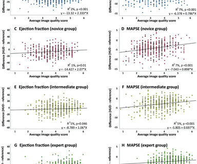
Non-experts using handheld ultrasound devices (HUDs) challenge the clinical yield. Mitral annular plane systolic excursion (MAPSE) reflects LV long-axis shortening. Background and objectives Echocardiography is the cornerstone of heart failure (HF) diagnosis, but expertise is limited.
Focusing on mitral and tricuspid valve diseases , Capstans treatment combines transcatheter implantation of a folded valve replacement with its X-ray and ultrasound-guided robot to align the low-profile implant with the beating heart valve. The post Capstans Robotic MR/TR Frontier Funding appeared first on Cardiac Wire.
Though there are several parameters for evaluation of left ventricular diastolic function by echocardiography, the most commonly used are the pulsed Doppler mitral E/A ratio and tissue Doppler mitral E/e’ ratio. Doppler interrogation of mitral valve is usually done from the apex through the apical four chamber view.
Mitral valve prolapse (MVP) is a common condition affecting approximately 3% of the population, typically with a benign clinical course. However, a small subset of patients (510%) may develop severe mitral re.
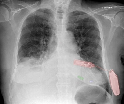
In the process, improving the ejection fraction and possibly reducing mitral regurgitation. Green: Micra leadless pacemaker; blue: WiSE-CRT system LV endocardial electrode; and red: WiSE-CRT system subcutaneous battery and ultrasound generator. Leadless Ultrasound-Based Cardiac Resynchronization System in Heart Failure.
Serial echocardiographic assessments are common in clinical cardiology, e.g., for timing of intervention in mitral and aortic regurgitation. When following patients with serial echocardiograms, each new measur.
We did a bedside cardiac ultrasound. The structure at the bottom that is moving is the mitral valve, with anterior and posterior leaflets. The ECG and ultrasound could not have been differentiated from acute plaque rupture with occlusion of the RCA. 3 points gets you an MI by Sgarbossa. This is as clear a STEMI as you can get.
During echocardiography, a transducer transmits the ultrasound beam towards the heart. Tracing in the lower part is tissue Doppler imaging from the medial mitral annulus. Opening and closing movements of the aortic and mitral valves are visible. Hence a basic knowledge is needed for all physicians and paramedics.
My opinion was that it was not a cath lab case, but I did suggest they do a bedside ultrasound to look for an anterior wall motion abnormality. I had not seen the cardiac ultrasounds at this time. I did not have more information at the time. To the ED providers, the patient denied CP, SOB, or drug use.
Blunt cardiac injury my result in : 1) Acute myocardial rupture with tamponade 2) Valve rupture (tricuspid, aortic, mitral) 3) Coronary thrombosis or dissection (and thus Acute MI) from direct coronary blunt injury 4) Dysrhythmias of all kinds. In the ED, ultrasound showed hemopericardium with tamponade.
During medical school, one of the classic bedside exam questions we get is how to differentiate the valve issues of aortic stenosis and mitral regurgitation, which produce similar but different murmurs when you listen with a stethoscope. “How can you tell the difference between aortic stenosis and mitral regurgitation at the bedside?”
He had diffuse crackles on exam and B-lines on chest ultrasound, and chest x-ray also confirmed pulmonary edema. Of course, papillary muscle rupture and mitral regurgitation should be on the differential here, as in this case , but it is not very likely when the BP is so high. Blood pressure was 215/124 and HR 115 (on metoprolol).
Here are a couple shots with strain, or "speckle tracking" on ED Echo: To, me these look like anterior wall motion abnormality, but I showed them to one of our ultrasound fellows who is very interested in this. If it were me, I would get values at the level of the mitral valve, papillary muscles, and apex (all in PSS axis).
Image courtesy: Philips christine.book Wed, 06/12/2024 - 14:07 June 12, 2024 — Royal Philips has announced its next-generation AI-enabled cardiovascular ultrasound platform to help speed up cardiac ultrasound analysis with proven AI technology and reduce the burden on echocardiography labs.
A bedside cardiac ultrasound was performed with a parasternal long axis view demonstrated below: There is a large pericardial effusion with collapse of the right ventricle during systole. The beat-to-beat variation in QRS amplitude and morphology is electrical alternans. Some say this looks like someone jumping on a trampoline.
To, me these look like anterior wall motion abnormality, but I showed them to one of our ultrasound fellows who is very interested in this. If it were me, I would get values at the level of the mitral valve, papillary muscles, and apex (all in PSS axis). hyperacute T-waves speckle 2 x4 from Stephen Smith on Vimeo.
For example, the mitral valve can be repaired with a catheter-based system. With the patient under general anesthesia, the device is delivered to the heart through a catheter, starting in the groin and guided by X-ray and ultrasound. The new system is designed specifically for the tricuspid valve’s position, location and shape.
Developed at Children’s National Hospital and detailed in the latest edition of the Journal of the American Heart Association , the new AI system combines the power of novel ultrasound probes with portable electronic devices installed with algorithms capable of diagnosing RHD on echocardiogram. Beginning in March, Craig Sable, M.D.
Santos Most Cited Article – Reduction in Hospitalization and Increase in Mortality Due to Cardiovascular Diseases during the COVID-19 Pandemic in Brazil Authors: Paulo Garcia Normando, José de Arimatéia Araujo-Filho, Gabriela de Alcântara Fonseca, Rodrigo Elton Ferreira Rodrigues, Victor Agripino Oliveira, Ludhmila Abrahão Hajjar, André Luiz (..)
A bedside POC cardiac ultrasound was done: Findings: Decreased left ventricular systolic function. Mild to moderate mitral regurgitation. A cutoff of 1200 pg/ml for patients with a normal eGFR is very specific for heart failure. The patient was given furosemide and admitted to the hospital. Biatrial enlargement, severe.
I suspect pulmonary edema, but we are not given information on presence of B-lines on bedside ultrasound, or CXR findings. The scan showed a bicuspid aortic valve with severe stenosis and coronary artery disease. Or I suspect that there is OMI simultaneous with another pathology. We certainly know that there is hypoxia.
We organize all of the trending information in your field so you don't have to. Join thousands of users and stay up to date on the latest articles your peers are reading.
You know about us, now we want to get to know you!
Let's personalize your content
Let's get even more personalized
We recognize your account from another site in our network, please click 'Send Email' below to continue with verifying your account and setting a password.


Let's personalize your content