This site uses cookies to improve your experience. To help us insure we adhere to various privacy regulations, please select your country/region of residence. If you do not select a country, we will assume you are from the United States. Select your Cookie Settings or view our Privacy Policy and Terms of Use.
Cookie Settings
Cookies and similar technologies are used on this website for proper function of the website, for tracking performance analytics and for marketing purposes. We and some of our third-party providers may use cookie data for various purposes. Please review the cookie settings below and choose your preference.
Used for the proper function of the website
Used for monitoring website traffic and interactions
Cookie Settings
Cookies and similar technologies are used on this website for proper function of the website, for tracking performance analytics and for marketing purposes. We and some of our third-party providers may use cookie data for various purposes. Please review the cookie settings below and choose your preference.
Strictly Necessary: Used for the proper function of the website
Performance/Analytics: Used for monitoring website traffic and interactions
Transcipt of video: Mild tricuspid regurgitation is often noted on echocadiogram reports and sometimes causes a little bit of worry and a lot of questions are asked on mild tricuspid regurgitation. What is this mild tricuspid regurgitation? And mild tricuspid regurgitation is just a small leak from the tricuspid valve.
Prognostic impact of severe tricuspid regurgitation (TR) in patients with atrial functional mitral regurgitation (AFMR). Abstract Aims Tricuspid regurgitation (TR) is often seen in patients with atrial functional mitral regurgitation (AFMR). The secondary endpoint was all-cause mortality. in severe AFMR.
In Ebstein’s anomaly, there is downward or apical displacement of posterior and septal tricuspid leaflets. The anterior leaflet is not displaced, but is elongated to meet the other leaflets, so that when it closes, a loud sound, tricuspid sound, is produced, which is called as the sail sound.
Both atria develop from a combination of the primitive atrium, sinus venous, and pulmonary veins.It However, underlying lesions such as hypertension, mitral valve disease, COPD, ASD, and TR greatly influence the degree of atrial enlargement. But, we rarely dispute it , & ask which atrium dilates more in AF ? Let us see few factors.
Objectives The estimation of systolic pulmonary artery pressure (sPAP) by transthoracic echocardiography (TTE) is challenging in patients with severe tricuspid regurgitation (TR). The two principal indications were TR (34.3%) and mitral regurgitation (32.2%). years old; male 56%).
Methods and Results This case report discusses a 65-year-old man who had previously undergone pulmonary vein isolation (PVI) and cavo-tricuspid isthmus ablation for atrial fibrillation before ASD closure, respectively. He developed atrial tachycardia (AT) and underwent catheter ablation.
D-Transposition of the great arteries (TGA) is a rare congenital heart defect where the pulmonary artery originates from the left ventricle (LV) and the aorta from the right ventricle (RV).
Though there are several parameters for evaluation of left ventricular diastolic function by echocardiography, the most commonly used are the pulsed Doppler mitral E/A ratio and tissue Doppler mitral E/e’ ratio. Doppler interrogation of mitral valve is usually done from the apex through the apical four chamber view.
Results Correction of severe AS by TAVR significantly reduced the proportion of patients suffering from concurrent severe mitral regurgitation (from 9.29% to 3.64%, p value: 0.0015). Moreover, pulmonary artery pressures were ameliorated (estimated systolic pulmonary artery pressure: from 47.2±15.8 ±15.8 ±15.1
The tricuspid valve is the right atrioventricular valve. The bicuspid or mitral valve is the left atrioventricular valve is. The pulmonary semilunar valve is between the right ventricle and the pulmonary trunk. Like the heart chambers, there are four heart valves between each of the chambers.
To confirm the efficacy and safety of extra pulmonary vein (PV) ablation, patients were categorized into two groups: those undergoing pulmonary vein isolation (PVI) alone and those receiving additional ablation. Methods Data from early commercial use across seven European centers were collected in a registry.
The RFCs were much more successful at classifying murmurs from the pulmonary and tricuspid valves (AUROC = 0.83 and 0.78, respectively) when compared with the aortic and mitral valves (AUROC = 0.72 All RFC models were evaluated using the area under their receiver operating characteristic curves (AUROCs). and 0.65, respectively).
Tracing in the lower part is tissue Doppler imaging from the medial mitral annulus. Opening and closing movements of the aortic and mitral valves are visible. The aorta, right ventricular outflow tract and pulmonary artery up to its bifurcation is imaged in the upward angulation shown in the left panel.
We are blessed with 4 heart valves – 2 on the left side which are known as the mitral and aortic valves and 2 on the right side – the tricuspid and pulmonary valves.
ABSTRACT Background The impact of tricuspid regurgitation (TR) on the outcomes of pulmonary vein isolation (PVI) for atrial fibrillation (AF) remains unclear.
Background Significant secondary mitral regurgitation (SMR) is known to be associated with worse prognosis. However, data focusing specifically on moderate SMR and associated risk factors are lacking. After matching, NYHA class and SMR aetiology remained associated with both outcomes (for both: log rank p<0.050).
Program Designations Access and Publications (A&P) 1 Participant User File (PUF) 2 Task Force on Funded Research (TFR) 3 Special Projects 4 Adult Cardiac Surgery Database Lead Author Title Publication Date William Keeling 2 National Trends in Emergency Coronary Artery Bypass Grafting European Journal of Cardiothoracic Surgery October 2023 Jake (..)
Bernoulli equation (P=4V 2 ) gives the gradient of tricuspid regurgitation flow, which corresponds to the pressure difference between right ventricle and right atrium in systole. The early diastolic pulmonary regurgitation (PR) gradient (peak PR gradient) will give an estimate of mean pulmonary artery pressure.
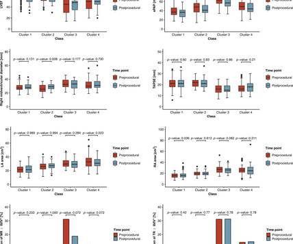
AF, atrial fibrillation; LAVI, left atrial volume index; RA, right atrial; RV, right ventricular; sPAP, systolic pulmonary artery pressure; SVI, stroke volume index; TR, tricuspid regurgitation. In addition, the TAPSE/systolic pulmonary artery pressure ratio (TAPSE/sPAP) was monitored as a measure of RVpulmonary arterial (PA) coupling.
We organize all of the trending information in your field so you don't have to. Join thousands of users and stay up to date on the latest articles your peers are reading.
You know about us, now we want to get to know you!
Let's personalize your content
Let's get even more personalized
We recognize your account from another site in our network, please click 'Send Email' below to continue with verifying your account and setting a password.


Let's personalize your content