This site uses cookies to improve your experience. To help us insure we adhere to various privacy regulations, please select your country/region of residence. If you do not select a country, we will assume you are from the United States. Select your Cookie Settings or view our Privacy Policy and Terms of Use.
Cookie Settings
Cookies and similar technologies are used on this website for proper function of the website, for tracking performance analytics and for marketing purposes. We and some of our third-party providers may use cookie data for various purposes. Please review the cookie settings below and choose your preference.
Used for the proper function of the website
Used for monitoring website traffic and interactions
Cookie Settings
Cookies and similar technologies are used on this website for proper function of the website, for tracking performance analytics and for marketing purposes. We and some of our third-party providers may use cookie data for various purposes. Please review the cookie settings below and choose your preference.
Strictly Necessary: Used for the proper function of the website
Performance/Analytics: Used for monitoring website traffic and interactions
Degenerative mitral valve disease is common. Up to a quarter of patients with degenerative mitral valve disease may be asymptomatic despite having severe valve regurgitation. Efforts should focus on establishing high-volume regional centres of excellence for mitral valve repair. Valve repair is the recommended intervention.
Transcript of the video: Closure line of aortic valve on M-Mode echocardiogram, is seen as central line, while in bicuspid aortic valve, it is an eccentric closure, nearer to one of the walls of the aorta. That is an important feature of bicuspid aortic valve on M-Mode echocardiogram. So this is a premature beat. is the normal range.
Objectives The association of pulmonary hypertension (PH) with the outcome after mitral transcatheter edge-to-edge repair (M-TEER) focusing on the new ESC/ERS guidelines definition for PH. Background PH is frequently found in patients with mitral regurgitation and is associated with lower survival rates. p<0.001).
In this study, we present the case of a patient who was diagnosed with severe acute onset heart failure secondary to torrential mitral regurgitation following COVID-19 pneumonia. This combination of ECMO and ECLS served as a bridge to open mitral valve replacement 6 days after presentation.
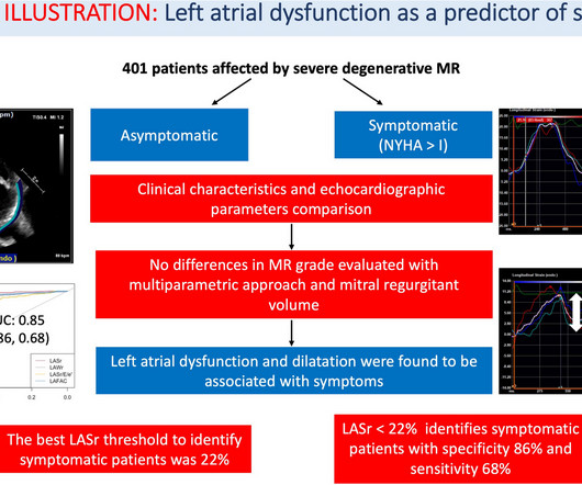
Left atrium (LA) is far from simply being a passive connection chamber between left ventricle and the pulmonary circulation. In patients affected by mitral regurgitation (MR) an impairment in LA compliance and.
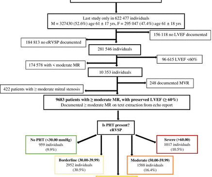
Objective Pulmonary hypertension (PHT) commonly coexists with significant mitral regurgitation (MR), but its prevalence and prognostic importance have not been well characterised. In a large cohort of adults with moderate or greater MR, we aimed to describe the prevalence and severity of PHT and assess its influence on outcomes.
AimOne of the philosophies of minimally invasive mitral surgery is to enhance recovery after surgery (ERAS). In this report, we aim to present institutional protocol for ERAS and its results in patients who underwent totally endoscopic mitral valve surgery (TEMVS).Patients 4) Rehabilitation; Physical and pulmonary rehabilitation. (5)
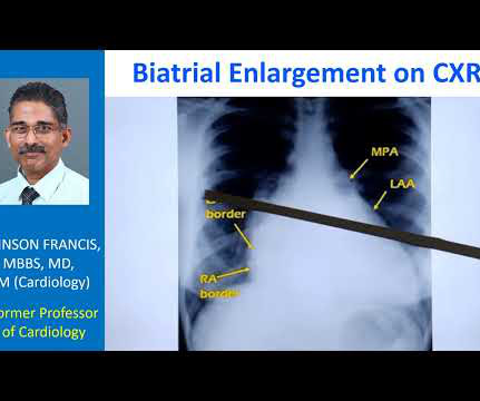
Both atria develop from a combination of the primitive atrium, sinus venous, and pulmonary veins.It However, underlying lesions such as hypertension, mitral valve disease, COPD, ASD, and TR greatly influence the degree of atrial enlargement. But, we rarely dispute it , & ask which atrium dilates more in AF ? Let us see few factors.
The interplay of MR and HFpEF: understanding increased pulmonary pressures and strategies for management. HFpEF, heart failure with preserved ejection fraction; MR, mitral valve regurgitation. Abstract Mitral regurgitation (MR) is highly prevalent among patients with heart failure and preserved ejection fraction (HFpEF).
This paper describes an instance of left ventricular bullet embolism from the pulmonary venous system following gunshot chest trauma. After 6 months, there were no signs or symptoms of cardiothoracic infection or evidence of mitral valve regurgitation in echocardiography.
Methods and Results This case report discusses a 65-year-old man who had previously undergone pulmonary vein isolation (PVI) and cavo-tricuspid isthmus ablation for atrial fibrillation before ASD closure, respectively. He developed atrial tachycardia (AT) and underwent catheter ablation.
Prognostic impact of severe tricuspid regurgitation (TR) in patients with atrial functional mitral regurgitation (AFMR). Abstract Aims Tricuspid regurgitation (TR) is often seen in patients with atrial functional mitral regurgitation (AFMR).
Though there are several parameters for evaluation of left ventricular diastolic function by echocardiography, the most commonly used are the pulsed Doppler mitral E/A ratio and tissue Doppler mitral E/e’ ratio. Doppler interrogation of mitral valve is usually done from the apex through the apical four chamber view.
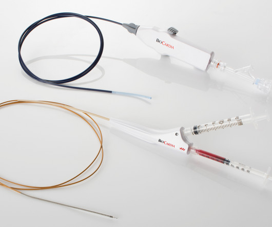
a company focused on cellular and cell-derived therapeutics for the treatment of cardiovascular and pulmonary diseases announced that the Unites States Patent Office has granted Patent No: 12,036,371 titled “Method of Accessing the Left Atrium with a Multi-Directional Steerable Catheter,” with a patent term that will expire in 2035.
He was in acute distress from pulmonary edema, with a BP of 180/110, pulse 110. He had diffuse crackles on exam and B-lines on chest ultrasound, and chest x-ray also confirmed pulmonary edema. The hypertension alone is the likely etiology of the pulmonary edema. He had no chest pain. Is this acute STEMI?
BACKGROUND:Patients with only moderate atrial secondary mitral regurgitation (asMR) frequently develop heart failure (HF). Although mild/moderate primary mitral regurgitation is compensated by left ventricular (LV) dilatation, the LV is not dilated in asMR. Circulation: Cardiovascular Imaging, Ahead of Print.
Clinical introduction A woman in her 30s, a case of rheumatic mitral stenosis status post balloon mitral valvuloplasty 15 years prior, presented to urgent care with palpitations and dyspnoea for 1 week. Echocardiography demonstrated severe calcific mitral stenosis with pulmonary hypertension.
This is the schematic diagram of the heart in which you can see right atrium, right ventricle, left atrium, left ventricle, aorta and pulmonary artery. Unlike the valves on the left side like the mitral and aortic, right sided valves can have some leak. Similarly, another right sided valve is the pulmonary valve.
Transcript of video: Hypoplastic Left Heart Syndrome is a very severe form of congenital heart disease, in which, the left ventricle, aorta and mitral and aortic valves are hypoplastic and valves may be atretic as well. A Gore-Tex tube is used and this maintains, this is a Blalock-Taussig shunt, which maintains pulmonary circulation.
Abstract Introduction Some previous studies have reported that a first-step ethanol infusion into the vein of Marshall (EIVOM) with touch-up radiofrequency (RF) ablation can facilitate mitral isthmus (MI) block and improves the ablation outcomes in persistent atrial fibrillation (PeAF) patients.
Objectives The estimation of systolic pulmonary artery pressure (sPAP) by transthoracic echocardiography (TTE) is challenging in patients with severe tricuspid regurgitation (TR). The two principal indications were TR (34.3%) and mitral regurgitation (32.2%). years old; male 56%).
We investigated whether estimated perimitral conduction time (E-PMCT), namely, twice the time between coronary sinus (CS) pacing and the ensuing wave-front collision at the opposite side of the mitral annulus, correlated with the cycle length of PMAT and could predict future PMAT.
D-Transposition of the great arteries (TGA) is a rare congenital heart defect where the pulmonary artery originates from the left ventricle (LV) and the aorta from the right ventricle (RV).
The bicuspid or mitral valve is the left atrioventricular valve is. The pulmonary semilunar valve is between the right ventricle and the pulmonary trunk. Several major arteries and veins include: The pulmonary artery transports blood with low levels of oxygen and high levels of carbon dioxide to the lungs.
Clinical introduction A woman in her mid-40s with a recent diagnosis of pulmonary embolism (on rivaroxaban) presented to the emergency room with dyspnea and fatigue. CT revealed stable pulmonary emboli compared with prior imaging, along with a new splenic infarct. Infectious workup was unrevealing.
1 PFA is conventionally used for pulmonary vein isolation (PVI), but interest has arisen in delivering linear lesions to the posterior wall2 and mitral isthmus.3 Pulsed field ablation (PFA) is a novel modality shown to be safe and efficient.1 3 The durability of these lesion sets has not been well evaluated.
The MitraClip procedure, is designed to reduce mitral regurgitation (MR) by approximating the mitral valve leaflets, can alter the direction or nature of residual MR, including potentially converting a central MR jet into an eccentric one. This could redirect the residual regurgitant flow. Implication of new onset eccentric jet 1.Eccentric
Cardiovascular neurocristopathy, i.e., cardiopathy and vasculopathy, associated with the NCC could occur in the form of (1) cardiac septation disorders, mainly the aortico-pulmonary septum; (2) great vessels and vascular disorders; (3) myocardial dysfunction; and (4) a combination of all three phenotypes.
Sudden breathlessness at night in those with known heart disease is usually due to collection of fluid in the lungs (pulmonary edema). During day time, when one is walking about, any extra fluid in the body tends to collect in the legs, due to the effect of gravity.
Among these, a fistula between the left anterior descending artery and the pulmonary artery is the rarest variant, comprising about 17% of all coronary artery fistula cases.Case:A 54-year-old male, with a known history of atrial fibrillation and hypertension, presented to our emergency department with non-rotatory dizziness.
Results Correction of severe AS by TAVR significantly reduced the proportion of patients suffering from concurrent severe mitral regurgitation (from 9.29% to 3.64%, p value: 0.0015). Moreover, pulmonary artery pressures were ameliorated (estimated systolic pulmonary artery pressure: from 47.2±15.8 ±15.8 ±15.1
We would like to express our gratitude to Dr. Bergonti for the valuable points of discussion regarding our manuscript Repeat pulmonary vein isolation and anterior line ablation using a novel point-by-point pulsed-field ablation system.1 1 We too have read his work on RF ablation of the anterior line with great interest.2
Publication date: Available online 21 November 2024 Source: The American Journal of Cardiology Author(s): Giulia Masiero, Federico Arturi, Elisa Boscolo Soramio, Luca Nai Fovino, Tommaso Fabris, Francesco Cardaioli, Andrea Panza, Giulia Lorenzoni, Massimo Napodano, Chiara Fraccaro, Giuseppe Tarantini
Normally, the main pulmonary artery segment will be concave and left atrial appendage region also will be not prominent. So that is why we see straightening of left border, typically heard of in mitral stenosis with left atrial enlargement and mild pulmonary hypertension. Those are not very clear in this picture.
Commonly employed empiric strategies for catheter ablation (CA) of refractory atrial fibrillation (AF) beyond pulmonary vein isolation (PVI) include posterior wall isolation (PWI), linear ablation involving left atrial (LA) roof and mitral lines, as well as targeting of areas of low voltage / myopathy.
This is the aortic valve in closed position and mitral valve also appears to be closed in position. From the images you do not know whether the mitral valve is really fully closed or almost about to be closed. You require multiple views to see from where the pulmonary arteries are arising. This could be a conus tissue.
Pulmonary vein isolation (PVI) is the cornerstone of atrial fibrillation (AF) ablation for which the left atrium (LA) is usually accessed by the antegrade femoral venous route and transseptal puncture. However, in rare cases, alternative routes must be used to overcome anatomical challenges (congenital or acquired).
This year's Boot Camp covered training in cardiopulmonary bypass skills, vessel anastomosis, diagnostic and therapeutic endoscopies, open pulmonary lobectomy, TAVR, and wire skills.
During repeat ablation, ULTC ablation lesions were assessed for electrical block, including segment-based assessment of pulmonary vein (PV) ablation lesions. Baseline patient and ULTC procedure characteristics were evaluated. Arrhythmia outcomes after repeat ablation were evaluated.
Abstract Objective To evaluate the progression of electrophysiological phenomena in atrial fibrillation (AF) and elucidate the association between the left atrial conduction velocity (LACV) and AF recurrence after pulmonary vein isolation. The left atrium was mapped using a 20-pole electrode catheter combined with the CARTO3 system.
We know a small ASD decompresses mitral stenosis, and the combination of ASD and MS, Lutembacher, is a well-known syndrome called Lutembacher. This is to create a small regulatory orifice in the IAS ( A complicated term for a small ASD ) to decompress the LA and reduce pulmonary congestive symptoms. JAMA Cardiol.
Tracing in the lower part is tissue Doppler imaging from the medial mitral annulus. Opening and closing movements of the aortic and mitral valves are visible. The aorta, right ventricular outflow tract and pulmonary artery up to its bifurcation is imaged in the upward angulation shown in the left panel.
While the first one may radiate to the axilla and base, but usually not into the neck, it does reflect both aortic outflow obstruction and mitral regurgitation in patients with a large gradient.
We organize all of the trending information in your field so you don't have to. Join thousands of users and stay up to date on the latest articles your peers are reading.
You know about us, now we want to get to know you!
Let's personalize your content
Let's get even more personalized
We recognize your account from another site in our network, please click 'Send Email' below to continue with verifying your account and setting a password.


Let's personalize your content