This site uses cookies to improve your experience. To help us insure we adhere to various privacy regulations, please select your country/region of residence. If you do not select a country, we will assume you are from the United States. Select your Cookie Settings or view our Privacy Policy and Terms of Use.
Cookie Settings
Cookies and similar technologies are used on this website for proper function of the website, for tracking performance analytics and for marketing purposes. We and some of our third-party providers may use cookie data for various purposes. Please review the cookie settings below and choose your preference.
Used for the proper function of the website
Used for monitoring website traffic and interactions
Cookie Settings
Cookies and similar technologies are used on this website for proper function of the website, for tracking performance analytics and for marketing purposes. We and some of our third-party providers may use cookie data for various purposes. Please review the cookie settings below and choose your preference.
Strictly Necessary: Used for the proper function of the website
Performance/Analytics: Used for monitoring website traffic and interactions
Though there are several parameters for evaluation of left ventricular diastolic function by echocardiography, the most commonly used are the pulsed Doppler mitral E/A ratio and tissue Doppler mitral E/e’ ratio. Doppler interrogation of mitral valve is usually done from the apex through the apical four chamber view.
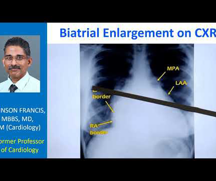
So that is why we see straightening of left border, typically heard of in mitral stenosis with left atrial enlargement and mild pulmonary hypertension. When there is gross pulmonary hypertension, instead of these being straight over here, it will form a bulge over here.
The post ectopic increase in the murmur is a hallmark of hypertrophic obstructive cardiomyopathy, which differentiates it clinically from mitral valve prolapse. Echocardiography in HCM Important echocardiographic features include mitral regurgitation and left ventricular outflow tract obstruction. in normotensives and more than 1.5
The cyanosis in Ebstein’s anomaly, is usually not due to pulmonary hypertension, but because tricuspid regurgitation jet is directed across the atrial septal defect. This distance, between the anterior mitral leaflet and septal tricuspid leaflet, is usually only about 5 mm during echocardiography.
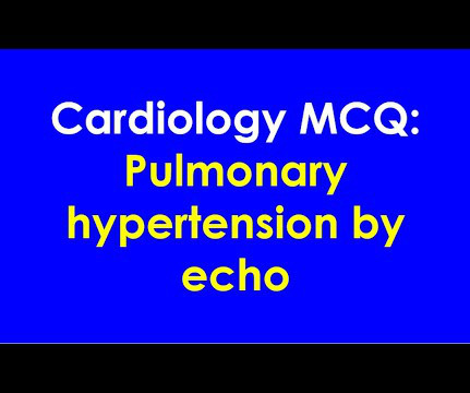
Most common method of assessment of pulmonary hypertension by Doppler echocardiography is by using: A: Forward velocity across the tricuspid valve B: Reverse velocity across the tricuspid valve C: Forward velocity across the pulmonary valve D: Reverse velocity across the mitral valve Correct answer: B: Reverse velocity across the tricuspid valve Reverse (..)
We organize all of the trending information in your field so you don't have to. Join thousands of users and stay up to date on the latest articles your peers are reading.
You know about us, now we want to get to know you!
Let's personalize your content
Let's get even more personalized
We recognize your account from another site in our network, please click 'Send Email' below to continue with verifying your account and setting a password.


Let's personalize your content