This site uses cookies to improve your experience. To help us insure we adhere to various privacy regulations, please select your country/region of residence. If you do not select a country, we will assume you are from the United States. Select your Cookie Settings or view our Privacy Policy and Terms of Use.
Cookie Settings
Cookies and similar technologies are used on this website for proper function of the website, for tracking performance analytics and for marketing purposes. We and some of our third-party providers may use cookie data for various purposes. Please review the cookie settings below and choose your preference.
Used for the proper function of the website
Used for monitoring website traffic and interactions
Cookie Settings
Cookies and similar technologies are used on this website for proper function of the website, for tracking performance analytics and for marketing purposes. We and some of our third-party providers may use cookie data for various purposes. Please review the cookie settings below and choose your preference.
Strictly Necessary: Used for the proper function of the website
Performance/Analytics: Used for monitoring website traffic and interactions
tim.hodson Wed, 04/16/2025 - 14:19 April 16, 2025 An artificial intelligence (AI) program trained to review images from a common medical test can detect early signs of tricuspid heart valve disease and may help doctors diagnose and treat patients sooner, according to research from the Smidt Heart Institute at Cedars-Sinai. Photo: Getty.
Transcipt of video: Mild tricuspid regurgitation is often noted on echocadiogram reports and sometimes causes a little bit of worry and a lot of questions are asked on mild tricuspid regurgitation. What is this mild tricuspid regurgitation? And mild tricuspid regurgitation is just a small leak from the tricuspid valve.
Echocardiograms using the robotic arm resulted in the same diagnosis as conventional in-person echocardiography in 98% of cases (papillary muscle level obstruction was missed in one case). tim.hodson Thu, 08/29/2024 - 11:39 Aug. 28, 2024 — New research presented at this year’s ESC Congress 2024 in London, UK (Aug. 30 – Sept.
Data from the studies demonstrated that AISAP CARDIO enables non-cardiologist physicians to interpret point-of-care echocardiograms just as well as expert cardiologists of the MGB echocardiography lab. James Hillis, MBBS, DPhil, director of Clinical Operations at Mass General Brigham AI.
A transthoracic echocardiogram (TTE) revealed a dilated LV with an EF of 20%, left atrial enlargement, severe mitral regurgitation, moderate tricuspid regurgitation, right ventricular systolic pressure of 42 mm HG, trivial pericardial effusion, akinesis of the inferior wall, and hypokinesis of the anterior wall.
From a total of 330 570 adult echocardiograms, 80 584 individuals were diagnosed with VHD and included in the final study population. In people aged 75 years, tricuspid and mitral regurgitation were the most prevalent VHD (21.1% Prevalence and correlates of VHD were assessed per each racial and ethnic group.
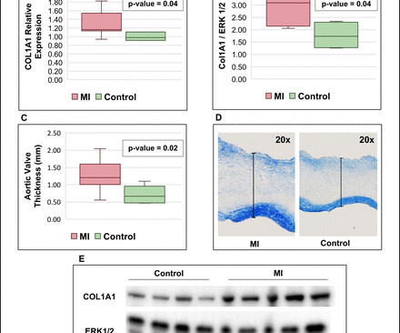
Background Myocardial infarction (MI) has been shown to induce fibrotic remodelling of the mitral and tricuspid valves. It is unknown whether MI also induces pathological remodelling of the aortic valve and alters aortic stenosis (AS) progression. Annualised progression rates of AS severity were compared between these 3 groups.
The image shown here is an animated 2 dimensional echocardiogram. This one is an older mode known as time-motion mode or M-Mode echocardiogram. Tracing in the lower part is tissue Doppler imaging from the medial mitral annulus. Opening and closing movements of the aortic and mitral valves are visible.
Blunt cardiac injury my result in : 1) Acute myocardial rupture with tamponade 2) Valve rupture (tricuspid, aortic, mitral) 3) Coronary thrombosis or dissection (and thus Acute MI) from direct coronary blunt injury 4) Dysrhythmias of all kinds. She was discharged to home feeling just fine.
Objective A substantial proportion of patients with rheumatic heart disease (RHD) have tricuspid regurgitation (TR). TR progression was defined either as worsening of TR degree from baseline to the last follow-up echocardiogram or severe TR at baseline that required surgery or died. and severe in 4.3%.
We organize all of the trending information in your field so you don't have to. Join thousands of users and stay up to date on the latest articles your peers are reading.
You know about us, now we want to get to know you!
Let's personalize your content
Let's get even more personalized
We recognize your account from another site in our network, please click 'Send Email' below to continue with verifying your account and setting a password.


Let's personalize your content