This site uses cookies to improve your experience. To help us insure we adhere to various privacy regulations, please select your country/region of residence. If you do not select a country, we will assume you are from the United States. Select your Cookie Settings or view our Privacy Policy and Terms of Use.
Cookie Settings
Cookies and similar technologies are used on this website for proper function of the website, for tracking performance analytics and for marketing purposes. We and some of our third-party providers may use cookie data for various purposes. Please review the cookie settings below and choose your preference.
Used for the proper function of the website
Used for monitoring website traffic and interactions
Cookie Settings
Cookies and similar technologies are used on this website for proper function of the website, for tracking performance analytics and for marketing purposes. We and some of our third-party providers may use cookie data for various purposes. Please review the cookie settings below and choose your preference.
Strictly Necessary: Used for the proper function of the website
Performance/Analytics: Used for monitoring website traffic and interactions
Transcript of the video: Now we will discuss echocardiogram in mitral valve prolapse. But, even though mitral valve prolapse can be detected echocardiographically in many cases, there may not be significant regurgitation and symptoms in many of them. So we will see some of the features of mitral regurgitation.
Transcript of the video: Closure line of aortic valve on M-Mode echocardiogram, is seen as central line, while in bicuspid aortic valve, it is an eccentric closure, nearer to one of the walls of the aorta. That is an important feature of bicuspid aortic valve on M-Mode echocardiogram. This is due an ectopic beat.
In this view, you can see that mitral leaflets are thickened. This is anterior mitral leaflet, thickened, and in the closed position of mitral valve, when there should be no flow to the left atrium, you are seeing a jet, a mosaic jet, which has been traced out. This is the mitral regurgitation jet.
There have been limited artificial intelligence studies published assessing the potential of machine learning to detect and analyze mitral regurgitation or to detect the presence of RHD on standard portable echocardiograms.Methods and ResultsWe used 511 echocardiograms in children, focusing on color Doppler images of the mitral valve.
Echocardiograms using the robotic arm resulted in the same diagnosis as conventional in-person echocardiography in 98% of cases (papillary muscle level obstruction was missed in one case). tim.hodson Thu, 08/29/2024 - 11:39 Aug. 28, 2024 — New research presented at this year’s ESC Congress 2024 in London, UK (Aug. 30 – Sept.
Transcript of the video: This is a still image from a colour Doppler echocardiogram, obtained from the apical five chamber view. Here, this is the forward flow through the mitral valve in diastole in red. Here also, the extent into the left atrium can be thought of as indicating severity of mitral regurgitation.
Cedars-Sinai and Smidt Heart Institute investigators developed a novel foundation model that integrates computer vision interpretation of echocardiogram images with natural language processing to augment cardiologists’ interpretation of echocardiograms. Image by Getty.
BACKGROUND:Many patients with atrial functional mitral regurgitation are not suitable candidates for surgery or transcatheter repair. Technical success and mitral regurgitation reduction from severe to none/trace were achieved in all cases. There were no cases of left ventricular outflow tract obstruction.
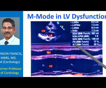
Transcript of the video: This is a still image of M-Mode Echocardiogram. Right ventricular outflow tract, left ventricle, left atrium, aorta, aortic valve, mitral valve. M-Mode is Time-Motion Mode. The horizontal axis is time. Vertical axis is distance from the transducer. In the inset you can see the two dimensional image.
Transcript of the video: This is a still image from a colour Doppler echocardiogram, obtained from the apical five chamber view. Here, this is the forward flow through the mitral valve in diastole in red. Here also, the extent into the left atrium can be thought of as indicating severity of mitral regurgitation.
BACKGROUND:Limited data exist regarding the impact of mitral annular calcification (MAC) on outcomes of transcatheter edge-to-edge repair for mitral regurgitation (MR).METHODS:We Circulation: Cardiovascular Interventions, Ahead of Print. with functional MR) who underwent an isolated, first-time intervention.
Serial echocardiographic assessments are common in clinical cardiology, e.g., for timing of intervention in mitral and aortic regurgitation. When following patients with serial echocardiograms, each new measur.
Data from the studies demonstrated that AISAP CARDIO enables non-cardiologist physicians to interpret point-of-care echocardiograms just as well as expert cardiologists of the MGB echocardiography lab. James Hillis, MBBS, DPhil, director of Clinical Operations at Mass General Brigham AI.
Unlike the valves on the left side like the mitral and aortic, right sided valves can have some leak. That is, mild mitral regurgitation and mild aortic regurgitation are less common. Mostly, they are detected on highly sensitive tests like echocardiogram. In echocardiogram, the Doppler beam can detect this small leak.
A transthoracic echocardiogram (TTE) revealed a dilated LV with an EF of 20%, left atrial enlargement, severe mitral regurgitation, moderate tricuspid regurgitation, right ventricular systolic pressure of 42 mm HG, trivial pericardial effusion, akinesis of the inferior wall, and hypokinesis of the anterior wall.
Inclusion criteria were: 1) who fulfilled the ESUS criteria based on NAVIGATE-ESUS trial, 2) who underwent transesophageal echocardiogram, and 3) admitted 7 days from the onset. RLS-ESUS showed higher frequency of anterior circulation lesion (RLS-ESUS vs. non RLS-ESUS; 84% vs. 71%, p = 0.035), slower early mitral inflow velocity (E) (51.6
Study participants (n=12 healthy volunteers, 40-66 years of age) underwent a focused baseline echocardiogram, which included atrial (A) and early (E) mitral valve inflow, peak tissue velocity (E’) at both the lateral and septal mitral annulus, and left ventricular ejection fraction (LVEF). cm/s (Δ 0.87
From a total of 330 570 adult echocardiograms, 80 584 individuals were diagnosed with VHD and included in the final study population. In people aged 75 years, tricuspid and mitral regurgitation were the most prevalent VHD (21.1% Prevalence and correlates of VHD were assessed per each racial and ethnic group. and 16.1%, respectively).
Similarly, for echocardiogram, what we would do usually is, first we do a clinical history evaluation, then physical examination, and after that only we proceed with echocardiography in our routine work. You can see the two dimensional sector imaging from an echocardiogram and I have marked out the aorta. This could be a conus tissue.
Residents also received instruction in mitral valve and aortic valve surgery, giving and receiving feedback in the operating room, and the importance of performing ablation. The best parts of the Boot Camp were learning the basics of CT surgery, the vast topics covered (transthoracic echocardiogram, lobectomy, etc.)
Introduction:Accurate detection of left atrial appendage (LAA) clot is critical in patients before Balloon Mitral Valvotomy (BMV). Transesophageal Echocardiogram (TEE) has traditionally been the gold standard for LAA clot detection. There is an elevated risk of thrombus formation and ischemic stroke in Rheumatic Heart Disease (RHD).
Medial mitral E/e' ratio >15 on transthoracic echocardiogram (TTE) defined elevated filling pressures. The AIECG assigned the LVDD grade (03). Inhospital and 1year mortality was evaluated, before and after multivariable adjustment.ResultsWe included 11 868 patients (median age 69.5
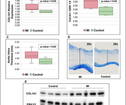
Background Myocardial infarction (MI) has been shown to induce fibrotic remodelling of the mitral and tricuspid valves. Methods Serial echocardiograms in human patients with AS were retrospectively analysed and compared between 3 groups: (1) acute MI at baseline (n=68), (2) prior history of MI (n=45) and (3) controls without MI (n=101).
The image shown here is an animated 2 dimensional echocardiogram. This one is an older mode known as time-motion mode or M-Mode echocardiogram. Tracing in the lower part is tissue Doppler imaging from the medial mitral annulus. Opening and closing movements of the aortic and mitral valves are visible.
Echocardiogram in parasternal long axis view shows dilated left ventricle, left atrium, aorta and a small portion of the right ventricle, which is usually the outflow region. Mitral valve leaflets seen in open position between the left ventricle and left atrium are thickened.
The post ectopic increase in the murmur is a hallmark of hypertrophic obstructive cardiomyopathy, which differentiates it clinically from mitral valve prolapse. Echocardiography in HCM Important echocardiographic features include mitral regurgitation and left ventricular outflow tract obstruction.
Blunt cardiac injury my result in : 1) Acute myocardial rupture with tamponade 2) Valve rupture (tricuspid, aortic, mitral) 3) Coronary thrombosis or dissection (and thus Acute MI) from direct coronary blunt injury 4) Dysrhythmias of all kinds. She was discharged to home feeling just fine.
Of course, papillary muscle rupture and mitral regurgitation should be on the differential here, as in this case , but it is not very likely when the BP is so high. Not all such ECGs represent anatomic aneurysms (on echo this is "diastolic dyskinesis"), but do generally represent an area of dense akinesis on echocardiogram.
Echocardiogram findings (pre-procedure) 1. Mitral valve calcification with mild regurgitation Laboratory data (pre-procedure) 1. LCx and RCA with luminal irregularities, but free of stenosis She was referred to cardiothoracic surgery, and underwent CABG x3 the following day. Normal LV/RV systolic function, EF 60-65 2. Troponin I 2.
If it were me, I would get values at the level of the mitral valve, papillary muscles, and apex (all in PSS axis). Even though the strain values are a little off in the graph (so is the posterior wall) it is still a value range (about -18) that would be considered non-ischemic by the cardiology literature, I believe.
Next day echocardiogram showed inferolateral hypokinesia with an EF of %45-50. On echocardiogram you will not see a "posterior" hypokinesia (will see "inferolateral") and, as in this case, LCx may not give the blood supply of basal inferior segment (formerly called "posterior"). The patient recovered well.
Developed at Children’s National Hospital and detailed in the latest edition of the Journal of the American Heart Association , the new AI system combines the power of novel ultrasound probes with portable electronic devices installed with algorithms capable of diagnosing RHD on echocardiogram. Beginning in March, Craig Sable, M.D.
TR progression was defined either as worsening of TR degree from baseline to the last follow-up echocardiogram or severe TR at baseline that required surgery or died. Methods A total of 645 patients with RHD were enrolled, mean age of 47±12 years, 85% female. Functional TR was graded as absent, mild, moderate or severe.
Shortly thereafter, a cardiac MRI was significant for probable HOCM based on septal hypertrophy, late gadolinium enhancement (LGE) with mid-wall fibrosis in the basal/mid inferolateral segments, and systolic anterior motion (SAM) of the mitral valve. Pre-stress echocardiogram revealed a sigmoid septum with septal wall thickness of 1.6
Mitral Valve Assessment Workshop: Corrado Fiore, MD (Italy), led a GE Healthcare-sponsored workshop on mitral valve assessment, featuring live echocardiogram scanning and hands-on sessions that guided participants from anatomical understanding to full diagnosis.
Echocardiogram is indicated (Correct) C. Start aspirin and Plavix Correct answer: (B) (B) Echocardiogram is indicated. While the first one may radiate to the axilla and base, but usually not into the neck, it does reflect both aortic outflow obstruction and mitral regurgitation in patients with a large gradient.
A 2D echocardiogram revealed an ejection fraction of 43%, hypokinesia of the anterior and intraventricular septum from base to apex, and severe mitral stenosis. Instead, the patient was treated with Aspirin 80 mg once daily, Clopidogrel 75 mg once daily, and Enoxaparin 0.4 ml subcutaneously once daily.
Later, he underwent a formal echocardiogram: Very severe left ventricular enlargement (LVED diameter 7.4 Mild to moderate mitral regurgitation. Severely decreased left ventricular systolic function with an estimated EF of 20-25%. No left ventricular wall motion abnormality identified.
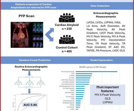
Machine learning applied to common measurements derived from routine echocardiogram studies can inform suspicion of CA. Methods We used 3603 echocardiogram studies from 636 patients at Cedars-Sinai Medical Center to train an RF model to predict CA from echocardiographic parameters.
More troponin values were measured at the cardiac center: 2327- 267 ng/L 0821- 355 ng/L 1108- 305 ng/L An echocardiogram on day three of the patients admission showed an ejection fraction of 46% with abnormal basal inferior and basal lateral segments, and severe aortic stenosis. No more EKGs were recorded during the patients admission.
We organize all of the trending information in your field so you don't have to. Join thousands of users and stay up to date on the latest articles your peers are reading.
You know about us, now we want to get to know you!
Let's personalize your content
Let's get even more personalized
We recognize your account from another site in our network, please click 'Send Email' below to continue with verifying your account and setting a password.


Let's personalize your content