This site uses cookies to improve your experience. To help us insure we adhere to various privacy regulations, please select your country/region of residence. If you do not select a country, we will assume you are from the United States. Select your Cookie Settings or view our Privacy Policy and Terms of Use.
Cookie Settings
Cookies and similar technologies are used on this website for proper function of the website, for tracking performance analytics and for marketing purposes. We and some of our third-party providers may use cookie data for various purposes. Please review the cookie settings below and choose your preference.
Used for the proper function of the website
Used for monitoring website traffic and interactions
Cookie Settings
Cookies and similar technologies are used on this website for proper function of the website, for tracking performance analytics and for marketing purposes. We and some of our third-party providers may use cookie data for various purposes. Please review the cookie settings below and choose your preference.
Strictly Necessary: Used for the proper function of the website
Performance/Analytics: Used for monitoring website traffic and interactions
Pathogeneses include connective tissue disorders, smooth muscle contraction disorders, and congenital heart disease, including bicuspid aortic valve, among others. Aortopathy is diagnosed commonly in children, from infancy through adolescence, primarily affecting the thoracic aorta, with variable involvement of the peripheral vasculature.
Circulation: CardiovascularImaging, Ahead of Print. Severe mitral regurgitation (MR) also causes a low-flow state, adding complexity to diagnosis and management.
Circulation: CardiovascularImaging, Ahead of Print. BACKGROUND:Patients with only moderate atrial secondary mitral regurgitation (asMR) frequently develop heart failure (HF). Although mild/moderate primary mitral regurgitation is compensated by left ventricular (LV) dilatation, the LV is not dilated in asMR.
Circulation: CardiovascularImaging, Ahead of Print. Mitral valve prolapse (MVP) affects 2% to 3% of the general population and is typically benign. However, a subset of patients may develop arrhythmic complications, including sudden cardiac arrest and sudden cardiac death.
However, if you freeze the ultrasound clip and scroll forwards and backwards to find a time during the clip where the patient’s mitral valve is open, you know the heart is filling, and is therefore in diastole. It is difficult to tell if there is collapse during diastole due to the patient’s tachycardia. J Am Soc Echocardiogr. 2013.06.023.
Circulation: CardiovascularImaging, Ahead of Print. BACKGROUND:A subset of patients with mitral valve prolapse (MVP), a highly heritable condition, experience sudden cardiac arrest (SCA) or sudden cardiac death (SCD). Each individual was categorized as having a normal mitral valve, MVP, or borderline MVP.
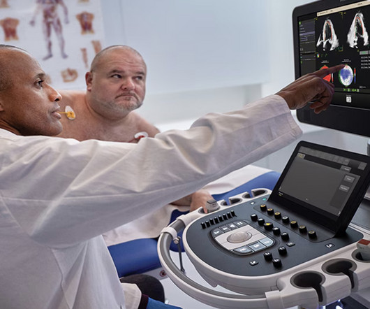
Image courtesy: Philips christine.book Wed, 06/12/2024 - 14:07 June 12, 2024 — Royal Philips has announced its next-generation AI-enabled cardiovascular ultrasound platform to help speed up cardiac ultrasound analysis with proven AI technology and reduce the burden on echocardiography labs.
The MHIF Imaging Core Lab, led by João Cavalcante, MD , director of the CardiovascularImaging Research Center at MHIF , provided advanced cardiac imaging analysis for the TRILUMINATE Pivotal trial. we are continuing to bring meaningful, life-enhancing benefits to patients with cardiovascular conditions."
demands thorough understanding of principles of Doppler echocardiography and also the hidden truths( ie, How we take liberty with the mighty Bernoulli equation for granted ) In spite of the number of imaging and doppler parameters we are able to gather ,still, we need to analyze them with reference to the clinical presentation. Reference 1.Zoghbi
We organize all of the trending information in your field so you don't have to. Join thousands of users and stay up to date on the latest articles your peers are reading.
You know about us, now we want to get to know you!
Let's personalize your content
Let's get even more personalized
We recognize your account from another site in our network, please click 'Send Email' below to continue with verifying your account and setting a password.


Let's personalize your content