This site uses cookies to improve your experience. To help us insure we adhere to various privacy regulations, please select your country/region of residence. If you do not select a country, we will assume you are from the United States. Select your Cookie Settings or view our Privacy Policy and Terms of Use.
Cookie Settings
Cookies and similar technologies are used on this website for proper function of the website, for tracking performance analytics and for marketing purposes. We and some of our third-party providers may use cookie data for various purposes. Please review the cookie settings below and choose your preference.
Used for the proper function of the website
Used for monitoring website traffic and interactions
Cookie Settings
Cookies and similar technologies are used on this website for proper function of the website, for tracking performance analytics and for marketing purposes. We and some of our third-party providers may use cookie data for various purposes. Please review the cookie settings below and choose your preference.
Strictly Necessary: Used for the proper function of the website
Performance/Analytics: Used for monitoring website traffic and interactions
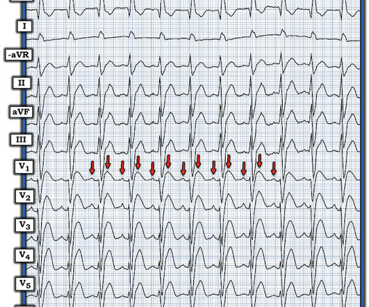
Initial assessment with electrocardiography revealed a regular narrow complex tachycardia with 2:1 atrioventricular (AV) relationship, no clear isoelectric baseline and positive P waves in lead V1 consistent with an atrialflutter of left atrial origin with a rapid ventricular rate (~160/min) ( figure 1A ).
edits by Meyers A woman in her 60s with a history of chronic atrial fibrillation on Eliquis, ESRD on hemodialysis, type-II diabetes mellitus, prior CVA, hypertension, and hyperlipidemia presented to the emergency department with multiple complaints after missing dialysis. They are flutter waves, and the rhythm is 2:1 atrialflutter.
male with pertinent past medical history including Atrial fibrillation, atrialflutter, cardiomyopathy, Pulmonary Embolism, and hypertension presented to the Emergency Department via ambulance for respiratory distress and tachycardia.
A fibrillatory wave that occurs at a rate of more than 600 beats per minute can cause fatigue in the long run, leading to atrial dilation. In all probability, this dilation is a form of atrial tachycardia and atrial cardiomyopathy. We know atrialflutters can be confined to one atrium.
For participants who slept just 5 hours per night, hypertension risk increased by 29%, depression risk increased by 64%, and anxiety risk increased by 46%. One last important finding regarding sleep duration was the J-shaped association between nightly sleep duration and the risk of hypertension, anxiety, and depression.
The rhythm is 2:1 atrialflutter. The flutter waves can conceal or mimic ischemic repolarization findings, but here I don't see any obvious findings of OMI or subendocardial ischemia. Chronic RVH is due to chronic pulmonary hypertension, and these patients are extremely difficult to manage when they are acutely ill.
A 50-something man with history only of alcohol abuse and hypertension (not on meds) presented with sudden left chest pain, sharp, radiating down left arm, cramping, that waxes and wanes but never goes completely away. 2 months later, he presented in pulmonary edema with atrialflutter and formal echo had EF 20% Why did this happen?
M Y A NSWER: In my experience, MAT is the 2nd-most commonly overlooked cardiac arrhythmia ( surpassed only by AtrialFlutter ). = Q UESTION : Why is M AT ( M ultifocal A trial T achycardia ) so commonly overlooked? How can you avoid overlooking this arrhythmia? Providers FORGET to “ U se t he O dds”.
He was hypertensive and tachycardic, with mildly increased work of breathing. The rhythm differential for narrow, regular, and tachycardic is sinus rhythm, SVT (encompassing AVNRT, AVRT, atrial tach, etc), and atrialflutter (another supraventricular rhythm which is usually considered separately from SVTs). If so, why?
Among all covariates, claims algorithms for covariates had sensitivities 85% for identifying diabetes, atrialflutter/fibrillation, and hypertension in MA and FFS. The kappa was higher in MA versus FFS for diabetes (P=0.03) and hypertension (P=0.025) but was lower in myocardial infarction (P<0.0001).
Methods The primary effectiveness endpoint (PEE) was 12-month freedom from documented atrial fibrillation/atrialflutter/atrial tachycardia plus freedom from acute procedural failure, nonstudy catheter failure, repeat ablation failure, direct current cardioversion (DCCV), and Class I/III antiarrhythmic drug (AAD) failure.
Edits by Meyers and Smith A man in his 70s with PMH of hypertension, hyperlipidemia, type 2 diabetes, CVA, dual-chamber Medtronic pacemaker, presented to the ED for evaluation of acute chest pain. LAFB, atrialflutter, anterolateral STEMI(+) OMI. Sent by Pete McKenna M.D. Triage ECG: What do you think? He expired 4 days later.
Figure-2: Colored arrows highlight flutter waves , with 2:1 AV conduction. Patient history: It turns out that todays patient is an 80-something year old man with longstanding hypertension and paroxysmal atrial fibrillation. Before each of these WCT episodes the atrial rate decreased.
We organize all of the trending information in your field so you don't have to. Join thousands of users and stay up to date on the latest articles your peers are reading.
You know about us, now we want to get to know you!
Let's personalize your content
Let's get even more personalized
We recognize your account from another site in our network, please click 'Send Email' below to continue with verifying your account and setting a password.


Let's personalize your content