This site uses cookies to improve your experience. To help us insure we adhere to various privacy regulations, please select your country/region of residence. If you do not select a country, we will assume you are from the United States. Select your Cookie Settings or view our Privacy Policy and Terms of Use.
Cookie Settings
Cookies and similar technologies are used on this website for proper function of the website, for tracking performance analytics and for marketing purposes. We and some of our third-party providers may use cookie data for various purposes. Please review the cookie settings below and choose your preference.
Used for the proper function of the website
Used for monitoring website traffic and interactions
Cookie Settings
Cookies and similar technologies are used on this website for proper function of the website, for tracking performance analytics and for marketing purposes. We and some of our third-party providers may use cookie data for various purposes. Please review the cookie settings below and choose your preference.
Strictly Necessary: Used for the proper function of the website
Performance/Analytics: Used for monitoring website traffic and interactions
male with pertinent past medical history including Atrial fibrillation, atrialflutter, cardiomyopathy, Pulmonary Embolism, and hypertension presented to the Emergency Department via ambulance for respiratory distress and tachycardia. Description : Regular Wide Complex Tachycardia at a rate of about 160.
She had a single chamber ICD/Pacemaker implanted several years prior due to ventricular tachycardia. The ECG was interpreted as showing atrialflutter with 2:1 conduction. Answer : The ECG above shows a regular wide complex tachycardia. The heart rate could be compatible with that of a 2:1 conducted atrialflutter.
Wide-complex tachycardia: VT or aberrant, or "other?" The patient had a history of paroxysmal atrial fibrillation and several cardioversions. A wide-complex tachycardia in an older patient must immediately suggest ventricular tachycardia. Instead, the rate of 150, plus the history of AF, suggested atrialflutter.
The 12-lead ECG and long lead II rhythm strip shown in Figure-1 — was obtained from a previously healthy, elderly woman who collapsed in the hospital parking lot. PEARL # 3: At this point — the most time-efficient step for solving today's rhythm will be to determine the nature of atrial activity.
His friend was able to get him into the truck and drive him to a nearby community hospital (non-PCI center). There is the appearance of STE in inferior leads II, III, and aVF (with STD in aVR), but this is entirely due to flutter waves which are only seen in those leads. Tachycardia and ST Elevation. Christmas Eve Special Gift!!
This strip was obtained: Apparent Wide Complex Tachycardia at a rate of 280 What do you think? To me, it was clearly atrialflutter with 1:1 conduction. The rate of 280 is just right for atrialflutter. The waves look like atrialflutter waves, NOT like a wide ventricular complex.
years, colchicine did not reduce a composite of emergency department visit, cardiovascular hospitalization, cardioversion, or repeat ablation (29 versus 25 per 100 patient-years; HR, 1.18 [95% CI, 0.69–1.99];P=0.55).CONCLUSIONS:Colchicine mg twice daily or placebo for 10 days. 11.53];P<0.001). During a median follow-up of 1.3
We see a regular tachycardia with a narrow QRS complex and no evidence of OMI or subendocardial ischemia. The differential of a regular narrow QRS tachycardia is sinus tachycardia, SVT, and atrialflutter with regular conduction. There are no P waves preceding the QRS complexes, and no clear flutter waves.
Electrical cardioversion may be recommended for you if you have certain types of arrhythmias, such as: Atrial fibrillation (AFib): This is the most common type of arrhythmia, and it can cause symptoms like dizziness, fatigue, and difficulty breathing. Atrialflutter: This is a rapid but regular heart rhythm often progressing to AFib.
Here was his ED ECG: There is sinus tachycardia (rate about 114) with nonspecific ST-T abnormalities. There is a large peaked P-wave in lead II (right atrial enlargement) There is left axis deviation consistent with left anterior fascicular block. The patient was given furosemide and admitted to the hospital. So what is it?
Introduction:Supraventricular tachycardia (SVT) is common and poorly tolerated in patients who have undergone Fontan procedure. During the 5-year follow up period, 13 (59%) patients with follow up had cardiovascular (CV) hospitalization and 1 patient died. Recurrence rates after catheter ablation in these patients are high.
2** Furthermore, the primary effectiveness endpoint (PEE) of acute pulmonary vein isolation and 12-month freedom from atrial arrhythmia recurrence (AF, AtrialTachycardia, or AtrialFlutter) was 75.6%. iii] The study reported a low fluoroscopy time of 7.8 iii] The study reported a low fluoroscopy time of 7.8
PurposeAtrial tachycardia is an uncommon supraventricular tachycardia in children. It is often drug-resistant and likely to occur concomitantly with tachycardia-induced cardiomyopathy, making radiofrequency catheter ablation the preferred treatment. A total of 78.6% No postoperative complications occurred in any patient.
If the patient has Abnormal Vital Signs (fever, hypotension, tachycardia, or tachypnea, or hypoxemia), then these are the primary issue to address, as there is ongoing pathology which must be identified. Clinical predictors of cardiac syncope at initial evaluation in patients referred urgently to general hospital: the EGSYS score.
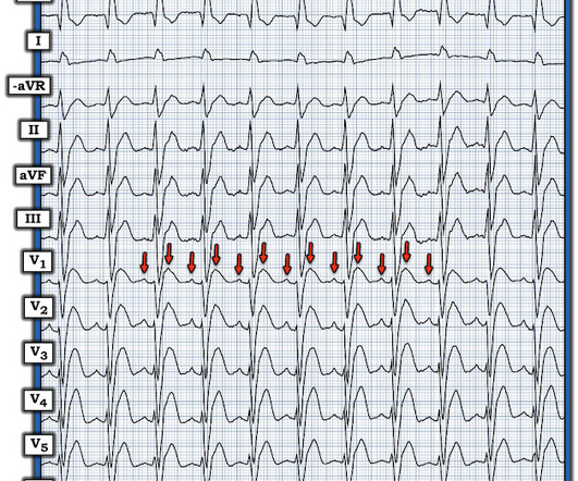
Figure-1: While at first glance the rhythm in Figure-1 might be mistaken for sinus tachycardia in fact, this is not the rhythm. Instead there is 2:1 atrial activity that is best seen in lead V1 ( See Figure-2 ). However, having noticed the 2:1 atrial activity in lead V1 I was not at all convinced that the episode was VT.
ABSTRACT Typical atrialflutter (AFL), defined as cavotricuspid isthmus (CTI)-dependent macro-re-entrant atrialtachycardia, often causes debilitating symptoms, and is associated with increased incidence of atrial fibrillation, stroke, heart failure, and death.
Smith comments : Wide complex tachycardia. The differential diagnosis of WCT is: 1) Sinus tachycardia with "aberrancy" (in this case RBBB and LAFB), but there are no P-waves and the QRS morphology is not typical of simple RBBB/LAFB. Also, if the rate is constant, not wavering up and down, it is highly unlikely to be sinus tachycardia.
We organize all of the trending information in your field so you don't have to. Join thousands of users and stay up to date on the latest articles your peers are reading.
You know about us, now we want to get to know you!
Let's personalize your content
Let's get even more personalized
We recognize your account from another site in our network, please click 'Send Email' below to continue with verifying your account and setting a password.


Let's personalize your content