This site uses cookies to improve your experience. To help us insure we adhere to various privacy regulations, please select your country/region of residence. If you do not select a country, we will assume you are from the United States. Select your Cookie Settings or view our Privacy Policy and Terms of Use.
Cookie Settings
Cookies and similar technologies are used on this website for proper function of the website, for tracking performance analytics and for marketing purposes. We and some of our third-party providers may use cookie data for various purposes. Please review the cookie settings below and choose your preference.
Used for the proper function of the website
Used for monitoring website traffic and interactions
Cookie Settings
Cookies and similar technologies are used on this website for proper function of the website, for tracking performance analytics and for marketing purposes. We and some of our third-party providers may use cookie data for various purposes. Please review the cookie settings below and choose your preference.
Strictly Necessary: Used for the proper function of the website
Performance/Analytics: Used for monitoring website traffic and interactions
In the study, HeartBeam AI with VCG demonstrated a 28% improvement over single-lead ECG in the detection of atrialflutter cases (sensitivity of 91.0% for single-lead ECG) without sacrificing the ability to identify those individuals without atrialflutter (specificity of 98.7% for VCG vs. 71.2% for VCG vs. 96.9%
It is unclear how deep learning applied to VCG compares to physicians (EPs) for atrialflutter (AFL) detection. The representation of ECG vectors in X, Y, and Z axes in a vectorcardiogram (VCG) has shown diagnostic promise beyond single lead ECG (SL) analysis, with implications for novel ECG acquisition technologies.
The positive F wave in lead V1 changed during entrainment from the right atrial appendage (RAA) during typical atrialflutter (AFL). Abstract Introduction Typical atrialflutter (AFL) is a macroreentrant tachycardia in which intracardiac conduction rotates counterclockwise around the tricuspid annulus.
In a world where technology reigns supreme, one of the most profound tools in medicine remains the irreplaceable electrocardiogram (ECG). AFIB/AFL – atrial fibrillation or atrialflutter episodes. An abnormal electrocardiogram can mean many things.
Here is an example where the computer failed to diagnose atrial fibrillation, with disastrous consequences: Computer often fails to diagnose atrial fibrillation in ventricular paced rhythm, and that can be catastrophic Smith SW et al. IJC Heart and Vasculature 25(2019). Poon et al. sensitivity and 98.9%
Electrocardiogram (ECG) abnormalities can be found in almost all patients, with Wolff–Parkinson–White (WPW) syndrome being the most common. However, many patients may not present the typical presentation, especially in the early stage. She denied any family history of cardiovascular disease or sudden death.
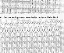
LAFB, atrialflutter, anterolateral STEMI(+) OMI. Fragmentation and artifact ( and possibly already in the inferior leads, the AtrialFlutter pointed out by Dr. Meyers that became obvious in the repeat ECG ) combine to make assessment of ST-T wave changes on many of the leads in ECG #1 difficult.
Abnormal Electrocardiogram (ECG): Defined (San Fran syncope rule) as any new changes when compared to the last ECG or presence of non-sinus rhythm. Results : Electrocardiograms (99%), telemetry (95%), cardiac enzymes (95%), and head computed tomography (CT) (63%) were the most frequently obtained tests.
We organize all of the trending information in your field so you don't have to. Join thousands of users and stay up to date on the latest articles your peers are reading.
You know about us, now we want to get to know you!
Let's personalize your content
Let's get even more personalized
We recognize your account from another site in our network, please click 'Send Email' below to continue with verifying your account and setting a password.


Let's personalize your content