Remove Atrial Flutter Remove Chest Pain Remove Ischemia
article thumbnail

Chest pain with anterior ST depression: look what happens if you use posterior leads.

Dr. Smith's ECG Blog

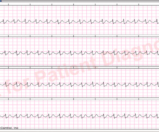
Written by Jesse McLaren A 65 year old with a history of atrial flutter, CABG and end-stage renal disease on dialysis presented with 3 days of fluctuating chest pain, which was ongoing at triage. The first ECG was labeled “anterior subendocardial ischemia”, but subendocardial ischemia does not localize.

article thumbnail

A man in his 70s with acute chest pain and paced rhythm.

Dr. Smith's ECG Blog

Edits by Meyers and Smith A man in his 70s with PMH of hypertension, hyperlipidemia, type 2 diabetes, CVA, dual-chamber Medtronic pacemaker, presented to the ED for evaluation of acute chest pain. LAFB, atrial flutter, anterolateral STEMI(+) OMI. EKG shown here: LAFB with no clear signs of OMI or ischemia.

article thumbnail

Medics were criticized for not activating the cath lab

Dr. Smith's ECG Blog

This is the prehospital ECG from an 81 year old man with acute chest pain. Here I put arrows: Arrows shows slow atrial flutter waves. And — it may be that this 81-year old man's "chest pain" may also disappear as soon as normal sinus rhythm is reestablished. The medics did NOT activate the cath lab.

article thumbnail

A 40-something presented after attempted prehospital resuscitation with persistent Ventricular Fibrillation

Dr. Smith's ECG Blog

My interpretation was: RBBB with hyperacute T-waves in V4-V6 that are all but diagnostic of LAD occlusion vs. post ROSC ischemia. Regional wall motion abnormality--apical anterior, mid anteroseptal, apical septal, and apical inferior akinesis. The patient had a volatile clinical course but awoke neuro intact.

article thumbnail

Very Fast Very Wide Complex Tachycardia

Dr. Smith's ECG Blog

A male in his 60's called 911 for dizziness and chest pain, onset with exertion. If it is slow Atrial flutter with 1:1 conduction, it should slow the conduction and reveal the flutter waves. Some of the ST depression is due to flutter waves, but there is also significant subendocardial "demand" ischemia.

article thumbnail

Ischemic ST depression maximal in V1-V4 (vs. V5-V6), even if less than 0.1 millivolt, is specific for Occlusion Myocardial Infarction (vs. subendocardial non-occlusive ischemia)

Dr. Smith's ECG Blog

A 50-something man with history only of alcohol abuse and hypertension (not on meds) presented with sudden left chest pain, sharp, radiating down left arm, cramping, that waxes and wanes but never goes completely away. 2 months later, he presented in pulmonary edema with atrial flutter and formal echo had EF 20% Why did this happen?

article thumbnail

A Middle-Aged Man with Chest pain, Hypotension and Tachycardia

Dr. Smith's ECG Blog

In the evening, a middle-aged man complained of chest pain at the nursing home. His chest pain was vague. He mentioned "cancer" and "chest". Leads II and aVF appear to have flutter waves. I diagnosed atrial flutter with 2:1 conduction. He was awake, with a pulse of 130 and BP of 50/30.