This site uses cookies to improve your experience. To help us insure we adhere to various privacy regulations, please select your country/region of residence. If you do not select a country, we will assume you are from the United States. Select your Cookie Settings or view our Privacy Policy and Terms of Use.
Cookie Settings
Cookies and similar technologies are used on this website for proper function of the website, for tracking performance analytics and for marketing purposes. We and some of our third-party providers may use cookie data for various purposes. Please review the cookie settings below and choose your preference.
Used for the proper function of the website
Used for monitoring website traffic and interactions
Cookie Settings
Cookies and similar technologies are used on this website for proper function of the website, for tracking performance analytics and for marketing purposes. We and some of our third-party providers may use cookie data for various purposes. Please review the cookie settings below and choose your preference.
Strictly Necessary: Used for the proper function of the website
Performance/Analytics: Used for monitoring website traffic and interactions
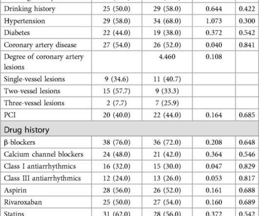
ObjectiveThis study aimed to investigate the effects of Enhanced External Counterpulsation (EECP) on anxiety and depression in patients with Paroxysmal AtrialFibrillation (PAF).MethodsA MethodsA cohort of 100 patients diagnosed with PAF at the Fuzhou First Hospital between January 2023 and June 2024 were enrolled in this study.
male with pertinent past medical history including Atrialfibrillation, atrial flutter, cardiomyopathy, Pulmonary Embolism, and hypertension presented to the Emergency Department via ambulance for respiratory distress and tachycardia. Bedside ultrasound showed volume depletion and no pulmonary edema. SVT with aberrancy?
Atrial cardiomyopathy is closely associated with atrialfibrillation (AF), and some patients exhibit no dysfunction at rest but demonstrate evident changes in left atrial (LA) function and LA volume during exe.
Titled "Real-world Data Affirms Safety and Effectiveness of Low/Zero Fluoroscopy AtrialFibrillation Ablation," the study was presented as a late-breaker at the 29th Annual International AF Symposium. The updated workflow indicates that direct imaging guidance, such as ultrasound, may be used as an alternative to fluoroscopy. "As
Several predictors of atrialfibrillation (AF) onset in patients with hypertrophic cardiomyopathy (HCM) have been proposed, however, all of them showed limited accuracy. This study aims to assess the role of n.
Bedside ED ultrasound showed exceedingly poor global LV function, and no B lines. So it must be atrialfibrillation. Answer : it is irregularly irregular and the initial part of the QRS is fast, so this is atrialfibrillation with Left Bundle Branch Block (LBBB). Here is the initial ED ECG. What do you think?
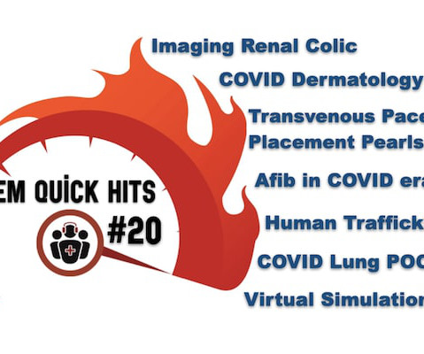
The post EM Quick Hits 20 Imaging Renal Colic, Human Trafficking, AtrialFibrillation During COVID, Transvenous Pacemaker Placement, COVID Lung POCUS, COVID Derm, Virtual Simulation appeared first on Emergency Medicine Cases.
Her Apple Watch suddenly told her that she is in atrialfibrillation. Patients with healthy AV nodes who are not on AV nodal blockers and who are not hyperkalemic should have a rapid ventricular response if they have paroxysmal Atrialfibrillation. Exam was completely normal except for an irregular heart rate.
Methods Using endoscopy, endoscopic ultrasound, and electrogastrography before and after PVI, esophageal and periesophageal injury (mucosal lesions, food retention, periesophageal edema, or vagal nerve injury) were assessed following PFA and radiofrequency (RF)- or cryoballoon (CB)-PVI.
Intracardiac echocardiography is increasingly utilized for atrialfibrillation (AF) ablation to perform transseptal puncture, monitor for complications and to define left atrial anatomy.
Our AI-enabled portfolio, including our Command Center Software Platform, Edison True PACS , and Venue Family ultrasound systems with Caption Guidance , is designed to directly address these issues. "At GE HealthCare, we understand the critical challenges healthcare providers face, from staffing shortages to complex workflows.
He arrived in the ED and had an immediate bedside cardiac ultrasound while this ECG was being recorded. The bedside ultrasound (video not available) reportedly showed only a slightly reduced LV function. It is dangerous in WPW with atrialfibrillation. Here is the ECG: What do you think?
This condition is known as atrialfibrillation, in which the upper chambers of the heart generate very fast irregular signals but fail to contract well. Ultrasound image of the heart – echocardiogram, showing fluid collection around the heart, marked as PE, short for pericardial effusion.
24 will focus on the following three current guideline updates: American College of Cardiology (ACC)/American Heart Association (AHA) Guidelines 2023 AtrialFibrillation Guideline - Pharmacology II: Strokes vs. Bleeds, What Do the Guidelines Tell Us About Practical Management in A-fib? The Guidelines Sessions at ACC.24
Adenosine is only unsafe in atrialfibrillation with WPW , which presents with a very rapid rate, polymorphic QRS, and some very short RR intervals] Bedside echo showed poor LV fct. If it is antidromic AVNRT or AVRT (WPW, accessory pathway), it will convert. If it is flutter, it will reveal the underlying flutter waves.
At AMS Cardiology, we offer comprehensive cardiac care including services tailored to the specific needs of seniors such as: Echocardiography: This painless ultrasound uses sound waves to create images of your heart, allowing doctors to assess its structure and function.
Echocardiographic assessment of left ventricular diastolic function with special reference on diastolic function assessment in atrialfibrillation. In atrialfibrillation, the absence of atrial contraction and the A wave makes this assessment impossible. J Cardiovasc Ultrasound. J Cardiovasc Ultrasound.
At AMS Cardiology, we offer comprehensive cardiac care including services tailored to the specific needs of seniors such as: Echocardiography: This painless ultrasound uses sound waves to create images of your heart, allowing doctors to assess its structure and function.
What is the atrial activity? Or is it atrialfibrillation with complete AV block and junctional escape? Case continued A bedside ultrasound showed diminished LV EF and of course bradycardia. He appeared gray in color, with cool skin. Here is his ED ECG: There is bradycardia with a junctional escape.
Case submitted and written by Mazen El-Baba MD, with edits from Jesse McLaren and edits/comments by Smith and Grauer A 90-year old with a past medical history of atrialfibrillation, type-2 diabetes, hypertension, dyslipidemia, presented with acute onset chest/epigastric pain, nausea, and vomiting.
Here was his prehospital ECG, which I viewed immediately while the resident performed cardiac ultrasound: What do you think? Here is the cardiac ultrasound which the resident performed as I viewed the ECG: This shows a huge pericardial effusion. The patient converted to atrialfibrillation. Is is sinus?
Smith comment: This patient did not have a bedside ultrasound. Had one been done, it would have shown a feature that is apparent on this ultrasound (however, this patient's LV function would not be as good as in this clip): This is recorded with the LV on the right. In fact, bedside ultrasound might even find severe aortic stenosis.
A bedside cardiac ultrasound was performed with a parasternal long axis view demonstrated below: There is a large pericardial effusion with collapse of the right ventricle during systole. The beat-to-beat variation in QRS amplitude and morphology is electrical alternans. This patient is only pseudo-stable. She has already had syncope.
An international consensus statement on how to treat atrialfibrillation with catheter or surgical ablation has been published in EP Europace, a journal of the European Society of Cardiology (ESC), and was recently presented at EHRA 2024, a scientific congress held April 7-9 in Berlin, Germany.
Now, with Caption AI technology, clinicians using Vscan Air SL handheld ultrasound will have access to real-time, step-by-step guidance to capture diagnostic-quality images and automated ejection fraction estimation to help inform clinical decisions across cardiac settings. Of these, approximately 30 are AI-enabled ultrasound innovations.
Left untreated, tricuspid regurgitation can lead to atrialfibrillation , heart failure , kidney disease and even death. With the patient under general anesthesia, the device is delivered to the heart through a catheter, starting in the groin and guided by X-ray and ultrasound.
Here is his ECG: There is atrialfibrillation at a rate of 95. If detected early by ultrasound, the patient can be saved. Our own Dave Plummer of HCMC reported on survival of 2 of 6 patients with free wall myocardial rupture diagnosed by bedside ultrasound in the ED.(3) Exact pain history was difficult to ascertain.
Description There is atrialfibrillation at a rate of 95. If detected early by ultrasound, the patient can be saved. Our own Dave Plummer of HCMC reported on survival of 2 of 6 patients with STEMI who had free wall myocardial rupture diagnosed by presence of hemopericardium on bedside ultrasound in the ED.(3)
The rhythm is atrialfibrillation. Cardiac Ultrasound may be a surprisingly easy way to help make the diagnosis Answer: pulmonary embolism. Now another, with ultrasound. Initial ROSC was obtained, during which this ECG was obtained: What do you think? The QRS complex is within normal limits. What is the Diagnosis?
Ultrasound – this is easily available, very portable and usually a very low risk investigation. The main reason is to look for the presence of atrialfibrillation which is associated with stagnation of blood within the heart with consequent clot formation. There are a variety of ways to look at these.
Bedside ultrasound showed no effusion and moderately decreased LV function, with B-lines of pulmonary edema. There is atrialfibrillation. Comments: STEMI with hypokalemia, especially with a long QT, puts the patient at very high risk of Torsades or Ventricular fibrillation (see many references, with abstracts, below).
A bedside POC cardiac ultrasound was done: Findings: Decreased left ventricular systolic function. A cutoff of 1200 pg/ml for patients with a normal eGFR is very specific for heart failure. A diagnostic NT-proBNP cutoff of 900 pg/mL has been suggested in adults 50-75 years of age in absence of renal failure."
Check : [vitals, SOB, Chest Pain, Ultrasound] If the patient has Abdominal Pain, Chest Pain, Dyspnea or Hypoxemia, Headache, Hypotension , then these should be considered the primary chief complaint (not syncope). Aortic Dissection, Valvular (especially Aortic Stenosis), Tamponade. Good History and Physical exam, including a.
I suspect pulmonary edema, but we are not given information on presence of B-lines on bedside ultrasound, or CXR findings. increasing stenosis, ischemia, volume changes, increased blood pressure, atrialfibrillation, etc.) Or I suspect that there is OMI simultaneous with another pathology. What other pathology is possible?
We organize all of the trending information in your field so you don't have to. Join thousands of users and stay up to date on the latest articles your peers are reading.
You know about us, now we want to get to know you!
Let's personalize your content
Let's get even more personalized
We recognize your account from another site in our network, please click 'Send Email' below to continue with verifying your account and setting a password.


Let's personalize your content