This site uses cookies to improve your experience. To help us insure we adhere to various privacy regulations, please select your country/region of residence. If you do not select a country, we will assume you are from the United States. Select your Cookie Settings or view our Privacy Policy and Terms of Use.
Cookie Settings
Cookies and similar technologies are used on this website for proper function of the website, for tracking performance analytics and for marketing purposes. We and some of our third-party providers may use cookie data for various purposes. Please review the cookie settings below and choose your preference.
Used for the proper function of the website
Used for monitoring website traffic and interactions
Cookie Settings
Cookies and similar technologies are used on this website for proper function of the website, for tracking performance analytics and for marketing purposes. We and some of our third-party providers may use cookie data for various purposes. Please review the cookie settings below and choose your preference.
Strictly Necessary: Used for the proper function of the website
Performance/Analytics: Used for monitoring website traffic and interactions
Radiofrequency ablation (RFA) is an important therapeutic modality for atrialfibrillation (AF), widely utilized in clinical practice due to its safety and significant efficacy. This case report describes a unique instance of a patient developing AEF following AF ablation, accompanied by ischemic stroke and myocardial infarction.
Early treatment of persistent AF can reduce the risk of blood clots, stroke, and heart failure, and may prevent the disease from becoming permanent. Unlike paroxysmal AF, which describes symptoms that last for seven days or fewer, persistent AF is a sustained arrhythmia that lasts for more than a week 1. The company now anticipates U.S.
The competing risk of non-stroke mortality may limit the potential benefit of stroke prophylaxis therapy in patients with atrialfibrillation and/or atrialflutter (AF).
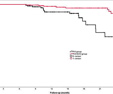
Background The value of empirical superior vena cava isolation (SVCI) following pulmonary vein isolation (PVI) to improve the efficacy of radiofrequency catheter ablation (RFCA) for paroxysmal atrialfibrillation (PAF) remains controversial.
A fully upright P-wave is typical atrial activity of atrialflutter as seen in V1. See these example cases of upright P-waves: Case Continued Thus, I was all but certain that this was atrialflutter. If it is flutter, it will reveal the underlying flutter waves. We want to avoid a stroke.
It is atrialflutter with 2:1 conduction. There are clear flutter waves in lead II across the bottom. In V1, there are upright waves that appear to be P-waves but are not: they are atrial waves and it is typical for atrialflutter waves to be upright in V1, whereas sinus P-waves are biphasic in V1.
67-year-old male has a planned PVI with RFA for atrialfibrillation involving bilateral wide area circumferential ablation, left carina line, posterior left atrial linear box isolation. He also underwent separate CTI ablation for atrialflutter. He was discharged the next day on colchicine and omeprazole.
Here is the computer interpretation: ATRIALFIBRILLATION WITH RAPID VENTRICULAR RESPONSE WITH ABERRANT CONDUCTION OR VENTRICULAR PREMATURE COMPLEXES LEFT AXIS DEVIATION [QRS AXIS beyone -30] NONSPECIFIC ST and T-WAVE ABNORMALITY The over-reading physician confirmed this diagnosis, which is incorrect. It is not atrialfibrillation.
Continue reading to learn more about this procedure, its significance in treating atrialfibrillation, and what to expect during treatment. What is AtrialFibrillation? Before diving into electrical cardioversion, we should understand atrialfibrillation (AF). What Is Cardioversion?
The electrical impulse goes further to the atrioventricular (AV) node, whose role is to slow down the conduction from the atria to the ventricles long enough for atrial contraction to occur. This allows the atria to fill the ventricles and achieve the highest possible stroke volume. An abnormal electrocardiogram can mean many things.
She also has a hx of paroxysmal atrialfibrillation and is on oral anticoagulant treatment. The ECG was interpreted as showing atrialflutter with 2:1 conduction. The heart rate could be compatible with that of a 2:1 conducted atrialflutter. After atrial rhythm/SR was restored the patient slowly improved.
Re-entrant tachycardias (atrialflutter, PSVT, AVRT, VT) have constant regular heart rates, whereas sinus tachycardia will usually gradually change rate with differing conditions (for instance, after infusion of fluid and BP increase, sinus tach rate might decrease from 130 to 125, for instance). So there is a re-entrant rhythm.
ABSTRACT Introduction The safety and efficacy of paroxysmal atrialfibrillation (PAF) ablation with the HELIOSTAR multielectrode radiofrequency (RF) balloon catheter have been demonstrated in European studies; data from elsewhere are lacking. Central Illustration. Image is courtesy of Biosense Webster, Inc., All rights reserved.
to 1.45) for fatal or nonfatal stroke. Of the 67 patients who underwent targeted tests, suspected diagnoses were confirmed in 49 (73%) patients: aortic stenosis (n = 8, 1%), pulmonary embolism (n = 8, 1%), seizures/stroke (n = 30, 5%), and other diseases (n = 3). g/dL Hypotension (obviously!) to 3.80).
ABSTRACT Typical atrialflutter (AFL), defined as cavotricuspid isthmus (CTI)-dependent macro-re-entrant atrial tachycardia, often causes debilitating symptoms, and is associated with increased incidence of atrialfibrillation, stroke, heart failure, and death.
We organize all of the trending information in your field so you don't have to. Join thousands of users and stay up to date on the latest articles your peers are reading.
You know about us, now we want to get to know you!
Let's personalize your content
Let's get even more personalized
We recognize your account from another site in our network, please click 'Send Email' below to continue with verifying your account and setting a password.


Let's personalize your content