This site uses cookies to improve your experience. To help us insure we adhere to various privacy regulations, please select your country/region of residence. If you do not select a country, we will assume you are from the United States. Select your Cookie Settings or view our Privacy Policy and Terms of Use.
Cookie Settings
Cookies and similar technologies are used on this website for proper function of the website, for tracking performance analytics and for marketing purposes. We and some of our third-party providers may use cookie data for various purposes. Please review the cookie settings below and choose your preference.
Used for the proper function of the website
Used for monitoring website traffic and interactions
Cookie Settings
Cookies and similar technologies are used on this website for proper function of the website, for tracking performance analytics and for marketing purposes. We and some of our third-party providers may use cookie data for various purposes. Please review the cookie settings below and choose your preference.
Strictly Necessary: Used for the proper function of the website
Performance/Analytics: Used for monitoring website traffic and interactions
BackgroundPlaque progression (PP) is critical between subclinical atherosclerosis and plaque rupture. Journal of the American Heart Association, Ahead of Print. Small dense lowdensity lipoprotein cholesterol (sdLDLC) is considered as the most atherogenic lipoprotein.
BackgroundCardiovascular disease (CVD) is a leading cause of death in women with systemic lupus erythematosus (SLE) due to accelerated atherosclerosis that is not predicted by established CVD risk scores. Despite having subclinical atherosclerosis, 44.8%
We report a case of TRAD in the early postoperative period, which was successfully managed with intravascular ultrasound-assisted endovascular intervention.Case presentationA 38-year-old man underwent HLA-compatible living kidney transplantation. Most cases are managed by operative repair.
Bedside cardiac ultrasound with no obvious wall motion abnormalities. Thus, angiography may be fairly accurate in determining lumen size, but it will not detect the “volume” of atherosclerosis present. He had a previous ECG on file: Proving the findings are new The cath lab was activated. He was started on nitro gtt.
Introduction:It remains uncertain whether dietary supplementation of marine n-3 polyunsaturated fatty acids (PUFAs) improves atherosclerosis and lipoprotein subclasses in patients with type 2 diabetes (T2D). The primary outcome was the prevalence of carotid artery plaques assessed by ultrasound. day) or low-dose (1.5g/day)
This study aimed to evaluate atherosclerosis as comorbidity by measuring the carotid (bulb and common carotid artery) Carotid intima-media thickness in COPD-diagnosed patients and to evaluate the relationship.
This manuscript provides a comprehensive review of intracranial atherosclerosis (ICAS)related largevessel occlusion (ICASLVO) and its mimics, focusing on the challenges in diagnosis and the need for precise diagnostic methodologies, particularly in the context of endovascular therapy.
Carotid atherosclerosis (CAS) is a critical precursor to atherosclerotic cardiovascular disease and is closely associated with the development and progression of conditions such as stroke and poor prognosis. Stroke, Volume 56, Issue Suppl_1 , Page ATP288-ATP288, February 1, 2025. 2021-KY-1289-001).Results:Among respectively.
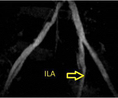
There is an area of dense white in the middle of the circle consistent with atherosclerosis. They too have dense white masses consistent with coronary atherosclerosis. . __ Here are some Images: The red circle shows the LAD coursing down the anterior interventricular sulcus. The green circle here shows a small section of the RCA.
Background:Lipoprotein a (Lp(a)) is known to be associated with coronary artery disease and carotid artery atherosclerosis. Carotid ultrasound results were divided into two groups based on the presence or absence of plaque. Lp(a) levels were categorized into two groups: below 50 mg/dl and 50 mg/dl or higher. 1692 (64.6%) were male.
Coronary Atherosclerosis There are multiple ways of describing coronary plaque: The degree of obstruction The composition of the plaque The thickness of the plaque wall (Cap) that separates the plaque from the inside of the blood vessel. One of the primary metrics used in studies of plaque regression is plaque volume or plaque area.
Before the procedure, patients should have an electrocardiogram (ECG) and echocardiogram (ultrasound of the heart) to check the heart’s rhythm and function. Lifetime risk of atrial fibrillation by race and socioeconomic status: ARIC Study (Atherosclerosis Risk in Communities). 2019;21(10):1468-1475. 7 Mou L, Norby FL, Chen LY, et al.
Background:Early detection of carotid artery (CA) conditions such as atherosclerosis and dissection are important as they represent major preventable causes of stroke. These patients had varying CA conditions (atherosclerosis, dissection, normal, etc.) Stroke, Volume 56, Issue Suppl_1 , Page AWMP107-AWMP107, February 1, 2025.
The fundamental characteristic of atherosclerosis is when a cholesterol particle becomes trapped in the artery wall. It is the inflammatory response to this particle retention that causes the formation of atherosclerosis 1. Lipoprotein particles entering the subintimal space causing atherosclerosis. You use an ultrasound.
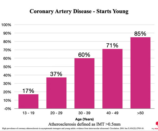
2 High prevalence of coronary atherosclerosis in asymptomatic teenagers and young adults: evidence from intravascular ultrasound. Subscribe now In Case You Missed Them: 1 Coronary artery calcium for the prediction of mortality in young adults <45 years old and elderly adults >75 years old. Eur Heart J. 2012 Dec;33(23):2955-62.
The problem is difficult to study because angiographic visualization of arteries is not perfect, and not all angiograms employ intravascular ultrasound (IVUS) to assess for unseen plaque or for plaque whose rupture and ulceration cannot be seen on angiogram. See "Mechanisms of acute coronary syndromes related to atherosclerosis".)
Doppler ultrasound was negative for DVT. Here, we describe a young patient with recurrent multi‐territory stroke, with the development of thrombocytopenia later in her course. In addition, it showed T2 hyperintensities in bilateral anterior and posterior watershed areas. Work‐up was significant for grade 3 PFO/intrapulmonary shunt.
This case was provided by Spencer Schwartz, an outstanding paramedic at Hennepin EMS who is on Hennepin EMS's specialized "P3" team, a team that receives extra training in advanced procedures such as RSI, thoracostomy, vasopressors, and prehospital ultrasound. See "Mechanisms of acute coronary syndromes related to atherosclerosis".)
In Ischaemic strokes, there is some sort of blockage either in the major vessels that take the blood (this is called large vessel atherosclerosis) to the brain or even in the smaller vessels (called small vessel occlusion). Ultrasound – this is easily available, very portable and usually a very low risk investigation.
The ways to tell for certain include intravascular ultrasound (to look for extra-luminal plaque with rupture) or "optical coherence tomography," something I am entirely unfamiliar with. The authors recommend using optical coherence tomography or intravascular ultrasound imaging in patients with evidence of nonobstructive CAD by angiogram.
Spontaneous coronary artery dissection Dissection of a coronary artery may occur in the context of atherosclerosis, or be iatrogennic during angiography or angioplasty. Often, intravascular ultrasound or intravascular optical coherence tomography is requeried to make the diagnosis. This case occurred 10+ years ago.
Atherosclerosis in the proximal segment of the vessel. Atherosclerosis, or plaque, is a progressive condition that occurs in stages over decades. Because the disease that is atherosclerosis is already very likely present in your coronary arteries as you read this. CT Coronary Angiogram. The more plaque, the higher the risk.
Methods We evaluated the associations between serum lipidomic profile and subclinical carotid atherosclerosis (SCA) in type 1 (T1D) and type 2 (T2D) diabetes, and in subjects without diabetes (controls) in a cross-sectional study.
Background:The presence of carotid plaque (CP) may serve as an indicator of panvascular atherosclerosis. We computed a Vascular Disease (VasD) score, integrating the presence of carotid plaque (CP) on carotid ultrasound, known coronary artery disease (CAD), and myocardial ischemia (MyI). Subsequently, patients were followed for 5.5
BackgroundObesity is accompanied by dysregulated inflammation, which can contribute to vasculometabolic complications including metabolic syndrome and atherosclerosis. Assessment of carotid artery atherosclerosis was performed with ultrasound. Journal of the American Heart Association, Ahead of Print.
While the exact mechanism underpinning that association is unclear, there is evidence that systemic inflammation contributes to atherosclerosis progression. One measure of subclinical atherosclerosis, carotid artery intima-media thickness (CIMT), is independently associated with ischemic stroke.
We organize all of the trending information in your field so you don't have to. Join thousands of users and stay up to date on the latest articles your peers are reading.
You know about us, now we want to get to know you!
Let's personalize your content
Let's get even more personalized
We recognize your account from another site in our network, please click 'Send Email' below to continue with verifying your account and setting a password.


Let's personalize your content