This site uses cookies to improve your experience. To help us insure we adhere to various privacy regulations, please select your country/region of residence. If you do not select a country, we will assume you are from the United States. Select your Cookie Settings or view our Privacy Policy and Terms of Use.
Cookie Settings
Cookies and similar technologies are used on this website for proper function of the website, for tracking performance analytics and for marketing purposes. We and some of our third-party providers may use cookie data for various purposes. Please review the cookie settings below and choose your preference.
Used for the proper function of the website
Used for monitoring website traffic and interactions
Cookie Settings
Cookies and similar technologies are used on this website for proper function of the website, for tracking performance analytics and for marketing purposes. We and some of our third-party providers may use cookie data for various purposes. Please review the cookie settings below and choose your preference.
Strictly Necessary: Used for the proper function of the website
Performance/Analytics: Used for monitoring website traffic and interactions
This confirms that the pain was ischemia and is now resovled. Thus, it has recently become generally accepted that most plaque ruptures resulting in myocardial infarction occur in plaques that narrow the lumen diameter by 40% of the arterial cross section may be involved by plaque. The i nitial hs troponin I returned 75%.
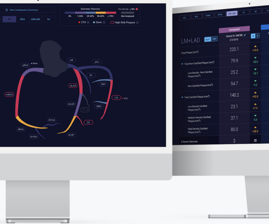
Food and Drug Administration ( FDA )-cleared Cleerly ISCHEMIA software device applied to a non-invasive coronary CT angiogram ( CCTA ) can be billed using the new Category I CPT code 75580. Prior studies have demonstrated the independent and incremental benefit of Cleerly ISCHEMIA applied to CCTA beyond traditional assessment.
This EKG is diagnostic of transmural ischemia of the inferior wall. The scan also showed “scattered coronary artery plaques”. __ Smith comment 1 : the appropriate management at this point is to lower the blood pressure (lower afterload, which increases myocardial oxygen demand). Lead I also shows reciprocal ST depression.
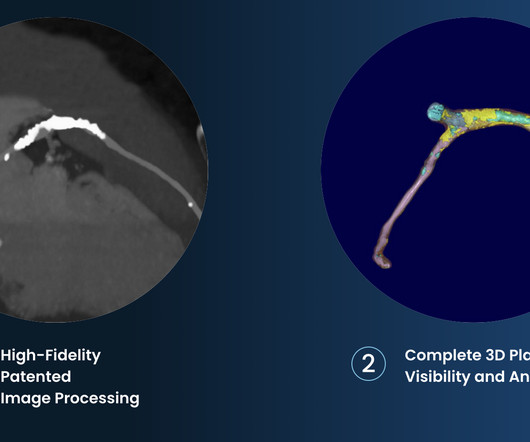
The company reports that its clinically-proven, AI-based digital care platform works with coronary computed tomography angiography (CCTA) imaging to help clinicians precisely identify and define atherosclerosis earlier, enabling them to provide personalized, life-saving treatment plans for all patients throughout their care continuum.
This suggests further severe ischemia. MINOCA may be due to: coronary spasm, coronary microvascular dysfunction, plaque disruption, spontaneous coronary thrombosis/emboli , and coronary dissection; myocardial disorders, including myocarditis, takotsubo cardiomyopathy, and other cardiomyopathies. And yet the arteries remain open.
The precordial STD persists in severity from V4-V6, rather than being maximal in V1-V4 (as in posterior OMI), and so the ECG overall best fits the subendocardial ischemia pattern (diffuse supply/demand mismatch). Meyers serves as a reminder of the important clinical entity known as diffuse subendocardial ischemia.
To prove there is no plaque rupture, you need to do intravascular ultrasound (IVUS). An angiogram is a "lumenogram;" most plaque is EXTRALUMINAL!! One of the most common is rupture of a non-obstructive plaque, with thrombus formation and OMI that spontaneously lyses and leaves a wide open artery. It can only be seen by IVUS.
T-wave inversion in V2 is inconsistent with early repol, and is typical of posterior ischemia. In addition, there is ST depression, diagnostic of ischemia, in V3-V6. Nevertheless, even young people have atherosclerosis and plaque rupture. mm of ST elevation in inferior leads. There should be no doubt that this is OMI.
This strongly suggests reperfusing RCA ischemia. Troponins, echocardiogram An echocardiogram showed inferobasilar hypokinesis, further supporting a diagnosis of regional ischemia , likely of the area supplied by the RCA. Here’s the angiogram of the RCA : No thrombus or plaque rupture in the RCA (or any coronary artery) was found.
This registry will aim to provide world-wide physicians the most accurate information on coronary plaque to improve cardiovascular risk prediction and support the selection of patient-specific treatment,” said Dr. De Cecco. The ultimate goal is to positively impact cardiovascular health globally with a reduction in cardiovascular events."
1 Atherosclerosis is a systemic disease that affects multiple vascular regions and is particularly severe in PAD patients, where up to 80 percent suffer from concurrent coronary artery disease (CAD), historically linked with a mortality rate exceeding 50 percent within five years. Journal of Vascular Surgery, Mar. 2024, [link].
Atherosclerotic cardiovascular disease (ASCVD), caused by plaque buildup in arterial walls, is one of the leading causes of disability and death worldwide.1,2 1,6 Until recently atherosclerosis has been thought of as the result of passive lipid accumulation in the vessel wall. 4 In the U.S.
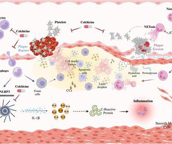
ACS may arise from the disruption of an atherosclerotic plaque, ultimately leading to acute ischemia and myocardial infarction. Colchicine exerts anti-inflammatory properties affecting both the myocardium and atherosclerotic plaque by modulating the activity of monocyte/macrophages, neutrophils, and platelets.
PAD is a serious, progressive cardiovascular disease primarily caused by a buildup of fatty plaque in the blood vessels, or atherosclerosis. This plaque narrows the blood vessels and reduces blood flow to the legs and feet, which may significantly impair physical function, walking performance and quality of life.
A CT CAC scan can only identify if there is calcified atherosclerosis, where it is and to what extent. A CTCA provides much more anatomical detail and can identify advanced plaque often missed by CT Coronary Artery Calcium Score scans alone. A CT CAC scan of 0 indicates no significant amount of calcified atherosclerosis.
Source: JAMA Cardiology) Patients with afib who survived an intracerebral haemorrhage had a very significant risk of cerebrovascular ischemia episodes and death in the following year, according to registry data. JACC: Asia) Lexaria Bioscience has announced that a CBD product beats a placebo in simulating acute pulmonary hypertension.
Poor blood supply Ischemia, or inadequate blood supply to the heart, is an abnormality that can be detected in an ECG test. Coronary artery disease Excessive cholesterol builds up plaque that blocks the arteries supplying blood to the heart. This condition is also called atherosclerosis.
Background:The presence of carotid plaque (CP) may serve as an indicator of panvascular atherosclerosis. We computed a Vascular Disease (VasD) score, integrating the presence of carotid plaque (CP) on carotid ultrasound, known coronary artery disease (CAD), and myocardial ischemia (MyI).
We organize all of the trending information in your field so you don't have to. Join thousands of users and stay up to date on the latest articles your peers are reading.
You know about us, now we want to get to know you!
Let's personalize your content
Let's get even more personalized
We recognize your account from another site in our network, please click 'Send Email' below to continue with verifying your account and setting a password.


Let's personalize your content