This site uses cookies to improve your experience. To help us insure we adhere to various privacy regulations, please select your country/region of residence. If you do not select a country, we will assume you are from the United States. Select your Cookie Settings or view our Privacy Policy and Terms of Use.
Cookie Settings
Cookies and similar technologies are used on this website for proper function of the website, for tracking performance analytics and for marketing purposes. We and some of our third-party providers may use cookie data for various purposes. Please review the cookie settings below and choose your preference.
Used for the proper function of the website
Used for monitoring website traffic and interactions
Cookie Settings
Cookies and similar technologies are used on this website for proper function of the website, for tracking performance analytics and for marketing purposes. We and some of our third-party providers may use cookie data for various purposes. Please review the cookie settings below and choose your preference.
Strictly Necessary: Used for the proper function of the website
Performance/Analytics: Used for monitoring website traffic and interactions
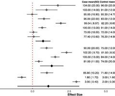
Our review study aimed to determine whether electrocardiogram (ECG) findings before PCI could serve as predictors for the occurrence of the no-reflow phenomenon. Result Sixteen eligible articles (1,473 cases and 4,264 controls) were included in this study. 2.38), P -value = 0.01), and Q-waves (OR (95% CI): 1.97 (1.01–2.94),
The article by Aslanger, Smith et al that is featured above in today’s post has just been published. The above-cited newly published article by Aslanger, Smith et al provides further support to the growing body of literature of why we should compel ourselves to do so. P.S.: Our September 3, 2020 post features Dr.
What is the specificity of exercise electrocardiogram stress testing (EST) in detecting ischemic substrate in patients with angina and nonobstructive coronary arteries (ANOCA)?
See these 2 articles Association between pre-hospital chest pain severity and myocardial injury in ST elevation myocardial infarction: A post-hoc analysis of the AVOID study Author links open overlay panel [link] 1 Background We sought to determine if an association exists between prehospital chest pain severity and markers of myocardial injury.
Persistent cardiac arrhythmias are readily amenable to detection by performing a standard electrocardiogram (ECG), but detection of transient (paroxysmal) arrhythmias has long been a significant cause of frustration to both doctors and patients. This article aims to provide a helpful overview for patients and doctors advising them.
Symptom-driven electrocardiogram (ECG) recording plays a significant role in the detection of post-ablation atrial fibrillation recurrence (AFR). The authors of an article published in Cardiovascular Innovations and Applications deployed a deep learning (DL)-based handheld device to facilitate symptom-driven monitoring.
A novel artificial intelligence (AI) model applied to a 12-lead electrocardiogram (ECG) image was able to identify individuals at elevated risk of heart failure (HF) across multinational cohorts without HF at baseline, according to a study published Jan. 13 in European Heart Journal.
Ryan Burch, RN, was the nurse caring for the patient, later sent me the same ECG, stating the following: "This ECG had people stumped and concerned but I read an article in www.ecgmedicaltraining.com (see below) about an artifact a few weeks prior which I thought looked similar and the suggestion was that a lead had been placed over an artery.
13 that Philips is correcting the Monitoring Service Application used with Mobile Cardiac Telemetry Monitoring as some electrocardiogram (ECG) events received by the application between July 2022 to July 2024 were not properly routed, which may have led to missing information or notifications.
The purpose of this article is to discuss possible ways of improving the quality of ECG signals in order to obtain more reliable results. Contractions of the heart muscle are caused by electrical impulses, which, when recorded on a graph, form an electrocardiogram. Why does the quality of the ECG signal matter?
Emergency department Code STEMI patients with initial electrocardiogram labeled ‘normal’ by computer interpretation: a 7-year retrospective review. For all cases, see the supplement from the online version of the article. This was just published in print in this month's Academic Emergency Medicine: McLaren, Meyers, Smith and Chartier.
What is the impact of a patient-initiated remote monitoring service including 12-lead electrocardiogram (ECG), vital sign data, and algorithmically supported clinical review on the risk of readmission following acute coronary syndrome (ACS)?
Similar promise is evident in the analysis of electrocardiograms, creating a new layer of interpretation. There is growing evidence that AI algorithms can match humans at echocardiography analysis, while also being able to extract subtle differences that the human eye cannot detect.
Comment A recently published article (1) found that a computer-interpreted “normal” ECG has a 99% NPV (95% CI: 97-99) for a clinically significant finding. According to 2 recent articles (2, 3), the computer misses one third of STEMI, but misses many more subtle MI that do not meet STEMI criteria. This one was not even so subtle!!!
Arterial pulse tapping artifact [link] This online article references the article below by Emre Aslanger, a great guy who occasionally corresponds with me about ECGs. Electromechanical association: a subtle electrocardiogram artifact. Figure-3: Page 475 from the Rowlands and Moore article that I reference above.
Cardiovascular consultation had been requested for all of the patients based on their primary clinical examination, vital signs, and electrocardiogram (ECG). In this cross-sectional study, 690 COVID-19 patients were included.
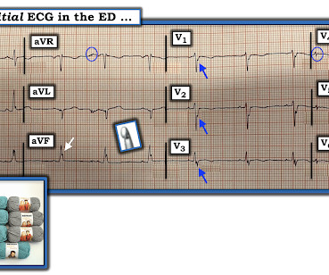
Here is an article I wrote: Updates on the ECG in ACS. Updates on the Electrocardiogram in Acute Coronary Syndromes. Electrocardiogram patterns in acute left main coronary artery occlusion. STE limited to aVR is due to diffuse subendocardial ischemia, but what of STE in both aVR and V1? If you want to understand aVR, read this.]
The electrocardiogram (ECG) revealed T-wave inversions on precordial leads. A 59-year-old woman presented to the emergency room with sudden onset of retrosternal thoracic pain following emotional stress. Her blood analyses demonstrated elevation of myocardial necrosis markers (peak of troponin I of 3.4
Many systems now refrain from showing computer "normal" ECGs to the busy emergency physicians at triage because of very poorly conceived articles that say that if the computer algorithm says "normal," the emergency physician should not be bothered. It is clearly missed by the conventional algorithm.
Here is the classic article on continuous 12-lead monitoring (in full text) showing that the ECG is a much more reliable indicator of re-occlusion than are symptoms. Here is another classic article. It is well documented with continuous 12-lead monitoring that acute re-occlusion is frequently asymptomatic. Patel DJ, et al.
Safety evaluation results showed no significant changes in the QRS wave or PR and QT intervals in rat lead II electrocardiograms. Safety evaluation results showed no significant changes in the QRS wave or PR and QT intervals in rat lead II electrocardiograms.
See the 5 articles below. Crochetage sign You can see many more images of the Crochetage sign here. N OTE : The term, “crochetage” in French is the noun ( = the act of crocheting ) — but this word has also been translated into English as meaning, “lock-picking” ( because lock-pickers often use a similar array of tools in their trade ).
The demographic, clinical, and electrocardiogram features of the patients in each group were compared. For symptomatic patients, the correlation between palpitations and PVC was further evaluated based on the temporal consistency of symptom onset and PVC occurrence. Results Of the 214 patients enrolled, 124(57.9%) experienced palpitations.
Normal QRS-T angle From this article: Ziegler R and Bloomfield DK. Yes, there are valuable articles from 50 years ago! A study of the normal QRS-T angle in the frontal plane. Journal of Electrocardiology 3(2):161-167; 1970.
The 12-lead electrocardiogram (ECG) and three-dimensional (3D) electroanatomical maps were analyzed. Methods This study enrolled 10 patients who underwent radiofrequency catheter ablation for a typical AFL. Electroanatomical mapping was performed both during typical AFL and entrainment from the right atrial appendage (RAA).
Methods and Results A 21-year-old female with supraventricular tachycardia (SVT) and pre-excitation on electrocardiogram (ECG) underwent electrophysiology study (EPS) confirming an AS-AP with anterograde and retrograde conduction. Ablation in the NCC achieved immediate and persistent anterograde conduction block.
FUJI trial, we assessed the differences in electrocardiogram (ECG) parameters during RV pacing between a delivery catheter system and a stylet system and their associations with the lead tip positions. In this subanalysis of the Mt.
In this article, we will explore the top 10 trends shaping the future of cardiovascular medicine, highlighting the innovative technologies and approaches that are revolutionizing the field. Traditional tools like stethoscopes, blood pressure gauges, and electrocardiograms (ECG) are fundamental for standard diagnostic practices.
This was just published in JAMA Internal Medicine: The de Winter Electrocardiogram Pattern Evolving From Hyperacute T Waves It reminded me that many believe, due to the assertions in the original de Winter's article, that de Winter's waves are stable.
Follow-up contained regular visits at our outpatient clinic at 1, 3, 6, and 12 months including 7-day Holter electrocardiograms. In addition to pulmonary vein isolation (PVI), ablation of DISPERS was performed aiming at homogenizing, dissecting, isolating, or connecting DISPERS areas to nonconducting anatomical structures. to 202.2 ± 21.6 ms
The ECG, as it turns out, is the best predictor , better the TMP grade because TMP measures microvascular patency, and the ECG measures cellular viability ( see this full text article and this abstract ). Clinical value of 12-lead electrocardiogram after successful reperfusion therapy for acute myocardial infarction.
Regional wall motion abnormality-distal septum anterior and apex Today, shortly after this case presented, I received in my inbasket from Journal Feed this article which promotes the idea that "normal" ECGs by computer do not need to be overread by a physician. Am J Emerg Med. 2022 Jan;51:384-387. doi: 10.1016/j.ajem.2021.11.023.
A deep neural network for 12-lead electrocardiogram interpretation outperforms a conventional algorithm, and its physician over-read, in the diagnosis of atrial fibrillation. IJC Heart and Vasculature 25(2019). Poon et al. sensitivity and 98.9%
When I teach important syncope ECG patterns to my residents, I have always had to find case reports and review articles to show them the rare epsilon waves in the past. 1211-1212 CrossRef View Record in Scopus Google Scholar 2 FI Marcus, W Zareba The electrocardiogram in right ventricular cardiomyopathy/dysplasia.
During this ceremony, we honored the best original articles, the most cited articles, and the most active reviewers of the journals ABC Cardiol, IJCS, ABC Imagem, and ABC HF. We thank all participants and collaborators who contributed to the success of our publications.
In this setting, family history of arrhythmia and being carrier of a pathogenic/likely pathogenic variant are the main risk factors for LV systolic dysfunction, while LV global longitudinal strain (LV-GLS) and Holter electrocardiogram (ECG) showed a relevant role in terms of prediction of LV systolic dysfunction and outcomes.
In the present article, we report the study protocol of the ResKriVer-TAVI trial. TIM will include a daily assessment of weight, blood pressure, a 2-channel electrocardiogram, peripheral capillary oxygen saturation, and a self-rated health status until admission for TAVI.
Safety of Computer Interpretation of Normal Triage Electrocardiograms. Here are two recent articles confirming this: a. The Comparison of Physician to Computer Interpreted Electrocardiograms on ST-elevation Myocardial Infarction Door-to-balloon Times. It is not yet available, but this is your way to get on the list.
You can find the answer and explanation at the end of this article. D) An electrocardiogram is most commonly normal in these patients. D) An electrocardiogram is most commonly normal in these patients. Cardiology Board Review Question A 48-year-old female with no known medical history presents with acute substernal chest pain.
Advanced arrhythmic substrate consisting of significant conduction abnormalities due to inflammation and fibrosis can be identified by specific electrocardiogram signs, such as fragmented QRS and ε wave. Methods The study population included consecutive 52 patients with CS and sustained VTA. Twenty-five out of 52 patients experienced ES.
At the bottom of the post, I have re-printed the section on aVR in my article on the ECG in ACS from the Canadian Journal of Cardiology: New Insights Into the Use of the 12-Lead Electrocardiogram for Diagnosing Acute Myocardial Infarction in the Emergency Department Case 1. Updates on the Electrocardiogram in Acute Coronary Syndromes.
This article discusses correction of the QT interval for rate. The article is written by Dr. Smith and Dr. Friedman. Essential Reading : Full text link: AHA/ACCF/HRS Recommendations for the Standardization and Interpretation of the Electrocardiogram, Part IV: The ST Segment, T and U Waves, and the QT Interval (full text link).
We organize all of the trending information in your field so you don't have to. Join thousands of users and stay up to date on the latest articles your peers are reading.
You know about us, now we want to get to know you!
Let's personalize your content
Let's get even more personalized
We recognize your account from another site in our network, please click 'Send Email' below to continue with verifying your account and setting a password.


Let's personalize your content