This site uses cookies to improve your experience. To help us insure we adhere to various privacy regulations, please select your country/region of residence. If you do not select a country, we will assume you are from the United States. Select your Cookie Settings or view our Privacy Policy and Terms of Use.
Cookie Settings
Cookies and similar technologies are used on this website for proper function of the website, for tracking performance analytics and for marketing purposes. We and some of our third-party providers may use cookie data for various purposes. Please review the cookie settings below and choose your preference.
Used for the proper function of the website
Used for monitoring website traffic and interactions
Cookie Settings
Cookies and similar technologies are used on this website for proper function of the website, for tracking performance analytics and for marketing purposes. We and some of our third-party providers may use cookie data for various purposes. Please review the cookie settings below and choose your preference.
Strictly Necessary: Used for the proper function of the website
Performance/Analytics: Used for monitoring website traffic and interactions
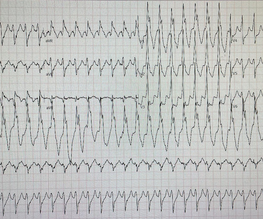
PEARL # 1: As I emphasize in ECG Blog #148 ( from where I took the tracing I show in Figure-3 ) — the BEST way to prove artifact — is to recognize persistence of an underlying spontaneous rhythm that is unaffected by any erratic or suspicious deflections that are seen. Figure-3: I've reproduced this tracing from ECG Blog #148 ( See text ). =
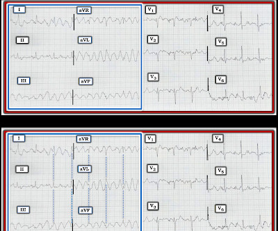
NOTE: The ECG in Figure-1 has been recorded at the usual 25mm/second speed — but with the Cabrera format ( Please see my Editorial Note near the top of the page in ECG Blog #365 for review of the basics of this recording system ). ECG Blog #185 — Review of the P s, Q s, 3 R Approach for systematic rhythm interpretation.
The most-popular HEART SISTERS posts from 2023 were all over the map - from 'Struggle Care' to sweating, hanging up that iconic Red Dress, or cardiac arrest on the toilet!
In the hope of dispelling continued dependence on millimeter-based STEMI criteria — we’ve published numerous cases in recent years in Dr. Smith’s ECG Blog of acute OMI ( O cclusion-based M yocardial I nfarction ) , in which patients have benefited from acute reperfusion despite not satisfying “STEMI criteria”.
. = I had a previous case of an adolescent with trauma and chest pain who also had AIVR: An adolescent with trauma, chest pain, and a wide complex rhythm From this blog post: "AIVR is NOT common in otherwise healthy children.
Readers of the Smith ECG Blog will probably recognize this a very subtle inferior OMI. The VT vs SVT with Aberrancy debate is beyond the scope of this particular blog post. Helpful tools to differentiate a WCT ECG include the Smith ECG Blog, and the Life in The Fast Lane blog. Here is the ECG after 200J.
As I discussed in detail in My Comment at the bottom of the page in the January 13, 2024 post in Dr. Smith's ECG Blog — pacemaker spikes tend to be a high frequency signal. The September 27, 2019 post — for the Rowlands & Moore article with the above-noted formulas for recognizing the “culprit” extremity.
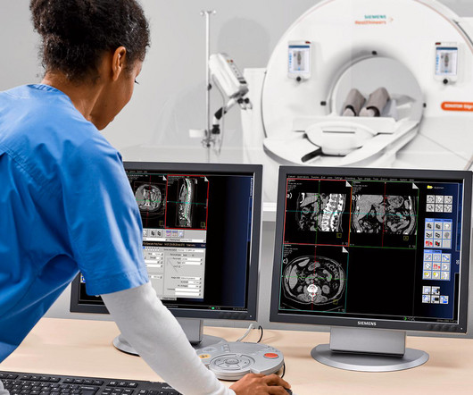
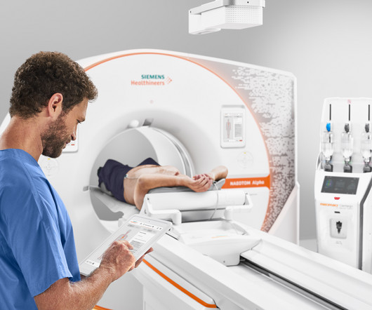
Insights into technology and what the future holds for MRI, CT and more were among our most viewed articles by far. As we reviewed our most popular publications of 2023, it became clear that many of you continue to rethink the limits of your profession. We’ll continue to bring you these insights and many more in 2024.
HYPERACUTE T-WAVES ARE EASILY RECOGNIZED BY THE QUEEN OF HEARTS HYPERACUTE T-WAVES DO NOT EVOLVE INTO ST ELEVATION -- THIS IS A MYTH Pendell and I are about to submit an article on ECG findings in total 100% LAD Occlusion (LAD OMI). For more on Precordial Swirl — See the October 15, 2022 post in Dr. Smith's ECG Blog ).
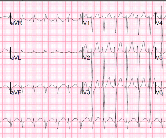
Readers of this blog can easily appreciate the hyperacute T waves in the precordium, clearest in V1-V4. We've highlighted a considerable number of acute RV MI cases in Dr. Smith's ECG Blog ( See the October 7, 2019 and May 10, 2024 posts , to name just two ). His ECG is shown below. Pretty obvious anterior current of injury.
Many systems now refrain from showing computer "normal" ECGs to the busy emergency physicians at triage because of very poorly conceived articles that say that if the computer algorithm says "normal," the emergency physician should not be bothered. We have shown many examples of this on this blog. No further follow up is available.
I described "Posterior Reperfusion T-waves" in this article. For more on ECG findings in pulmonary disease — Check out My Comment in the May 31, 2024 post in Dr. Smith's ECG Blog ). As often emphasized in Dr. Smith's ECG Blog — there is normally slight, upward sloping ST elevation in leads V2 and V3.
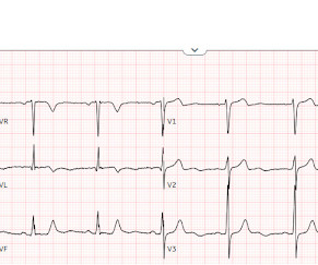
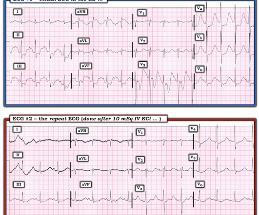
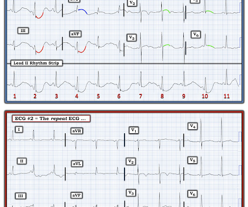
NOTE: The ECGs in today's case are recorded in the Cabrera Format ( See Dr. Grauer Comment in the October 26, 2020 post of Dr. Smith's ECG Blog for review on the Cabrera Format ). How would you manage this patient? Figure-1: The initial ECG in today's case. ( To improve visualization I've digitized the original ECG using PMcardio ).
As I have discussed the user-friendly and time-efficient criteria I favor for distinction between VT vs SVT rhythms ( with either preexisting BBB or aberrant conduction ) — I'll refer those interested in more detail to My Comments in the May 5, 2020 — the April 2, 2022 — and the February 14, 2022 posts in Dr. Smith's Blog.
When we reviewed our top articles of the year, it became clear that what resonated more than anything else were in-depth looks at technology, the cost of that technology, and the myriad other innovations changing the clinical conversation. Here are the top articles of 2024. 2024 was a year of innovation across the imaging spectrum.
Today's case is unique in that: i ) I believe it to be the 1st time we are publishing an example of this less commonly encountered form of lead reversal in Dr. Smith's ECG Blog; and , ii ) Because the patient's underlying baseline ECG abnormalities made recognition of this form of lead reversal that much more difficult to identify.
In 2022, a few key trends stuck out when reviewing Cassling's most popular articles. In this recap, check out the Top 10 articles of the year to discover what had the imaging world abuzz in 2022. The latest technology advances were top of mind, as was patient experience, staffing, and more.
Non-randomized trials show better outcomes (neurologic survival) using this device; see this article in Resuscitation: Head and Thorax Elevation during cardiopulmonary resuscitation using circulatory adjuncts is associated with improved survival. The patient had ROSC and maintained it. First — Some thoughts on the post -resuscitation ECG.
This is obviously unreliable data, as Dr. Smith’s Blog has published 51 cases of OMI with ECGs labeled ‘normal’ , 35 of which were identified by the Queen of Hearts – with 10 examples here. For all cases, see the supplement from the online version of the article. It should never have been published.
I have posted previous such cases, but in searching my own blog, I could not find them. Dr. Smith also referenced an article by Knotts et al, showing that even when diffuse subendocardial ischemia is due to coronary disease — that only a minority of patients had severe LMain coronary disease as the cause.
From webinars to informative articles, they are committed to spreading awareness and understanding. This committee will continue to be the driving force behind our efforts to influence public policy, raise awareness, and ensure that the CHD community’s voice is heard.
Smith : we have an article under review that shows that the variable most closely associated with the final diagnosis of "STEMI" vs. "Non-STEMI" was a door to balloon time less than, vs. greater than, 90 minutes. But because of the delayed reperfusion, the discharge was Non-STEMI.
NOTE: I reproduce in the ADDENDUM below ( in Figures-3 , - 4 and - 5 ) — the 3-page article by Rowlands and Moore ( J. As we have pointed out on multiple occasions ( See My Comment at the bottom of the page in the April 17, 2022 post in Dr. Smith's ECG Blog ) — lead V1,V2 malposition is surprisingly common in practice.
He has a great blog too: ECG Interpretation He is also well known on the Facebook EKG Club page , where you can learn tons about ECGs: Here is his response, with the first ECG labelled: Hello Steve & Avinash. It is commonly seen in the reperfusion setting. It appears to be benign in children as well (see references below).
See these 2 articles Association between pre-hospital chest pain severity and myocardial injury in ST elevation myocardial infarction: A post-hoc analysis of the AVOID study Author links open overlay panel [link] 1 Background We sought to determine if an association exists between prehospital chest pain severity and markers of myocardial injury.
26th August 2022 And so, after a great deal of faffing about, my article on cardiovascular disease ‘Assessing cardiovascular disease: looking beyond cholesterol’ has been made free to view. Writing an article for a medical journal is not that difficult. As for allowing the article to be open access … don’t go there.
Interpretation by the GE/Marquette 12 SL conventional algorithm Smith : This article, published this month (!) , tells us that we physicians do not need to even look at this ECG until the patient is placed in a room because the computer says it is normal: Validity of Computer-interpreted “Normal” and “Otherwise Normal” ECG in Emergency Department Triage (..)
Blog Introducing New Technology to an Institution jbagley Wed, 03/02/2022 - 11:15 Innovation and new technology are essential to the progress of any specialty. This article will discuss how to introduce new technology to an institution. Van Haren, MD, MsPH Career Development Blog Early Career Technology March 2, 2022 Robert M.
We've previously discussed the all-too-often ignored entity known as MINOCA ( = MI with N on- O bstructive C oronary A rteries ) — which we detailed in the November 30, 2022 post in Dr. Smith's ECG Blog ( See My Comment at the bottom of the page ). Don't assume that young people don't have OMI!!
These were read by our fantastic chief of radiology, Gopal Punjabi, who has his own blog on Spectral CT: [link] [link] Here is the image using Spectral CT : It is much more obvious with this technique! A slice at a slightly different level: Again, an area with absence of contrast enhancement (dark-, not light-colored).
Arterial pulse tapping artifact [link] This online article references the article below by Emre Aslanger, a great guy who occasionally corresponds with me about ECGs. NOTE: I reproduce below ( in Figures-3 , - 4 and - 5 ) — the 3-page article by Rowlands and Moore ( J. Aslanger E, Yalin K.
Ryan Burch, RN, was the nurse caring for the patient, later sent me the same ECG, stating the following: "This ECG had people stumped and concerned but I read an article in www.ecgmedicaltraining.com (see below) about an artifact a few weeks prior which I thought looked similar and the suggestion was that a lead had been placed over an artery.
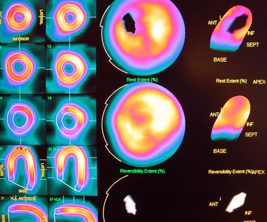
Therefore reading recent nuclear cardiology articles in journals accessible to you may be helpful. The post How to Effectively Prepare for the Nuclear Cardiology Boards appeared first on BoardVitals Blog. Recently in nuclear cardiology there has been a shift to emphasis on new hardware (i.e. solid state detectors), new software (i.e.
Articles on TCA More TCA ECGs from Dr. Smith's ECG Blog More on TCA overdose, with ECGs, from life in the fast lane. His delirium greatly improved and he was then able to follow commands. He had a prolonged stay in the ICU requiring days of bicarbonate. The tox screen only showed amitryptiline.
This article was originally published on AuntMinnie.com on August 21, 2023. Click here to view the original article. With all these variables on the table, it's no surprise that radiology technologists want tools at their disposal that will help them be efficient, effective, and at the forefront of the field.
For myself (and I suspect other readers of this blog), however, it affected my perception in exactly the opposite way – it immediately made me suspicious of misdiagnosis and wary of the dangers of diagnosing pericarditis (and therefore missing other diagnoses). There does not seem to be good evidence to answer this question, unfortunately.
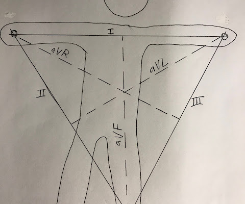
Normal QRS-T angle From this article: Ziegler R and Bloomfield DK. Yes, there are valuable articles from 50 years ago! A study of the normal QRS-T angle in the frontal plane. Journal of Electrocardiology 3(2):161-167; 1970.
data derived from Kashou et al article ECG interpretation proficiency of healthcare professionals In the previous study the difference in performance of ECG interpretation was mainly focused at cardiologists versus non cardiologist. The post ECG interpretation should and can improve appeared first on ECG-Excellence.
In the December 5, 2022 post of Dr. Smith's ECG Blog — We show 4 additional cases of this pulse-tap artifact. The September 27, 2019 post — for the Rowlands & Moore article with the above-noted formulas for recognizing the “culprit” extremity. This is no longer the case! Why are the smallest artifactual deflections GREEN ?
See this full text link to an article from JAMA on PCI in patients who present at 12 to 48 hours. Here is a link to a blog post with the formula , which we have recently validated and will publish. There is no question that this benefits from immediate PCI. As for thrombolytics, that is a bit riskier.
In the Marchik article, (assuming they defined it the same way, and the methods do not specify this), among patients with suspicion for PE, S1Q3T3 was found in 8.5% Finally , they found that S1Q3T3, precordial T-wave inversions V1-V4, and tachycardia were independent predictors of PE. What is an S1Q3T3? Very few studies define S1Q3T3.
A vast collection of webinars, CME activities, expert interviews, on-demand event recordings, news articles, drug pipelines, and many other resources are now grouped into areas of specialty focus.
We organize all of the trending information in your field so you don't have to. Join thousands of users and stay up to date on the latest articles your peers are reading.
You know about us, now we want to get to know you!
Let's personalize your content
Let's get even more personalized
We recognize your account from another site in our network, please click 'Send Email' below to continue with verifying your account and setting a password.


Let's personalize your content