This site uses cookies to improve your experience. To help us insure we adhere to various privacy regulations, please select your country/region of residence. If you do not select a country, we will assume you are from the United States. Select your Cookie Settings or view our Privacy Policy and Terms of Use.
Cookie Settings
Cookies and similar technologies are used on this website for proper function of the website, for tracking performance analytics and for marketing purposes. We and some of our third-party providers may use cookie data for various purposes. Please review the cookie settings below and choose your preference.
Used for the proper function of the website
Used for monitoring website traffic and interactions
Cookie Settings
Cookies and similar technologies are used on this website for proper function of the website, for tracking performance analytics and for marketing purposes. We and some of our third-party providers may use cookie data for various purposes. Please review the cookie settings below and choose your preference.
Strictly Necessary: Used for the proper function of the website
Performance/Analytics: Used for monitoring website traffic and interactions
Sympathetic overactivation and neuroinflammation in the paraventricular nucleus (PVN) are crucial factors in post-myocardial infarction (MI) cardiac remodeling and ventricular arrhythmias (VAs). Meanwhile, the cGAS-STING pathway has shown potential to ameliorate neuroinflammatory response.
Our previous study showed that light-emitting diode (LED) modulation of hypothalamic paraventricular nucleus (PVN), which is the control center of the sympathetic nervous system, might attenuate neuroinflammation in PVN and prevent ventricular arrhythmias (VAs) after myocardial infarction (MI).
BackgroundIschemic cardiomyopathy (ICM) is the end stage of ischemic heart disease, in which ventricular remodeling contributes to a fatal ventricular arrhythmia, worsens heart function and unfavorable outcomes, and is related to persistent chronic inflammation. Vagotomy was applied to suppress the cholinergic antiinflammatory pathway.
Bedside cardiac ultrasound showed moderately decreased LV function. (And of course Ken's comments at the bottom) An elderly obese woman with cardiomyopathy, Left bundle branch block, and chronic hypercapnea presented hypoxic with altered mental status. She was intubated. CT of the chest showed no pulmonary embolism but bibasilar infiltrates.
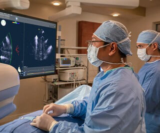
Due to the tethering of the LV tissue, 43% of MV Prolapse (MVP) subjects will develop ventricular arrhythmias that often remain undiagnosed or uncorrelated with MV disease presence. Electromechanical Wave Imaging (EWI) is a high frame rate ultrasound modality that noninvasively maps the electromechanical (EM) wave in all cardiac chambers.
He arrived in the ED and had an immediate bedside cardiac ultrasound while this ECG was being recorded. The bedside ultrasound (video not available) reportedly showed only a slightly reduced LV function. The patient was given 6mg, then 12 mg, of adenosine, without a change in the rhythm. Here is the ECG: What do you think?
a global leader in cardiac arrhythmia treatment and part of Johnson & Johnson MedTechi , revealed findings from a company-funded study of real-world data. The updated workflow indicates that direct imaging guidance, such as ultrasound, may be used as an alternative to fluoroscopy. "As
Euan Ashley (Stanford University) Safety Oversight of decentralized trials — Leanne Madre (Medable) Remote Arrhythmia Monitoring from an Academic Perspective — Greg Marcus (UCSF) Do Remote Heart-Monitoring Devices Work? REACT, LOOP etc.
Her bedside cardiac ultrasound was normal We decided to cardiovert her since the time of onset was very recent. The Role of Sinus Arrhythmia: I found it interesting to compare the long lead II rhythm strips in the 3 serial tracings from today’s case ( Figure-1 ). But when you see this, you should suspect that the AV node is not well.
A formal ultrasound later showed reasonably good LV function, and so he later received carvedilol and diltiazem, Unfortunately, those led to hypotension at 80/40 with a HR 40. PEARL #1: The most commonly overlooked arrhythmia is AFlutter ( A trial F lutter ). As a result — We can not rule out VT on the basis of this single ECG.
On intravascular ultrasound (IVUS), the mid RCA plaque was described as "cratered, inflamed, and bulky," and the OM plaque was described as "bulky with evidence of inflammation and probably ulceration." Additional findings: No ST elevation." They also documented "Reproducible chest tenderness."
Here was his prehospital ECG, which I viewed immediately while the resident performed cardiac ultrasound: What do you think? Here is the cardiac ultrasound which the resident performed as I viewed the ECG: This shows a huge pericardial effusion. Therefore, we performed ultrasound-guided pericardiocentesis. Is is sinus?
On arrival, the patient was in shock, was intubated, and had an immediate cardiac ultrasound. What does a heart look like on ultrasound when the EKG looks like that? Here you go: It's not the world's greatest cardiac ultrasound video, but it does appear to show poor function and low volume. They transported to the ED.
Smith comment: This patient did not have a bedside ultrasound. Had one been done, it would have shown a feature that is apparent on this ultrasound (however, this patient's LV function would not be as good as in this clip): This is recorded with the LV on the right. In fact, bedside ultrasound might even find severe aortic stenosis.
Other nonspecific findings may include P wave abnormalities, PR segment deviations, and atrial arrhythmias — though none of these findings are seen in a majority of patients. Electrical alternans is generally only seen with a large pericardial effusion. ii ) All 3 of the ECGs in this case manifest Schamroth’s S ign !
A bedside cardiac ultrasound was performed with a parasternal long axis view demonstrated below: There is a large pericardial effusion with collapse of the right ventricle during systole. Both ST segment and T wave alternans have been known to precede malignant ventricular arrhythmias. This patient is only pseudo-stable.
Regional wall motion abnormality-inferolateral (this is the formal ultrasound location of a posterior wall motion abnormality). Therefore, the rhythm in ECG #2 is a marked sinus arrhythmia — which was not clinically important, and which resolved within minutes by the time ECG #3 was recorded. Here is a case from a while back.
Widespread ST-depression with reciprocal aVR ST-elevation can be cause by: Heart rate related: tachyarrhythmia (e.g., A emergent cardiology consult can be helpful for equivocal cases. This finding does not alter the need to pursue emergent reperfusion, although it might suggest a poorer prognosis.”[3]
There are three mechanisms of arrhythmia: automatic, re-entry, and triggered. The most common triggered arrhythmia is Torsades de Pointes. It is a benign arrhythmia which requires no specific treatment. Possible mechanisms of ventricular arrhythmias elicited by ischemia followed by reperfusion. What is the rhythm?
However, he suddenly developed a series of malignant ventricular arrhythmias. Below are printouts of some of the arrhythmias recorded. This time, the arrhythmia did not spontaneously terminate — but rather degenerated to VFib, requiring defibrillation. The arrhythmia starts with a PVC having a short coupling interval.
It is also published in Heart Rhythm , the official journal of the HRS, Journal of Arrhythmia , the official journal of the APHRS, and Journal of Interventional Cardiac Electrophysiology , the official journal of the LAHRS. Antiarrhythmic drugs are advised for some patients to prevent arrhythmia recurrences early after the procedure.
My bedside ultrasound was of insufficient quality, but showed somewhat reduced overall EF, distended IVC without respiratory variation, no pericardial effusion, and diffuse bilateral B lines. == What do you think of her ECG? Of Note: This patient was hemodynamically stable without palpitations at the time ECG #1 was recorded.
Bedside ultrasound showed no effusion and moderately decreased LV function, with B-lines of pulmonary edema. IV administration of potassium is indicated when arrhythmias are present or hypokalemia is severe (potassium level of less than 2.5 malignant ventricular arrhythmias are present), rapid replacement of potassium is required.
A bedside ultrasound should be done to assess volume and other etiologies of tachycardia, but if no cause of type 2 MI is found, the cath lab should be activated NOW. Jenkins and Frick — I offer 3 additional examples of artifactual distortion ( excerpted from my ECG Blog ) — that resulted in arrhythmia misdiagnosis.
This was diagnosed by IVUS (intravascular ultrasound) as a ruptured plaque. Although there is no long lead rhythm strip — it is noteworthy that there is marked sinus arrhythmia in ECG #1. This finding is easy to overlook because of the lack of a long lead II … Perhaps this marked sinus arrhythmia reflects increased vagal tone?
Further ultrasound showed no B-lines (no pulmonary edema). WPW Cardiac arrhythmias ( especially AFib ). There is very little filling, and thus very poor stroke volume. The heart rate is too fast for this poor filling. Preload must be increased and the heart rate slowed in order to allow more LV filling.
A bedside POC cardiac ultrasound was done: Findings: Decreased left ventricular systolic function. By this I mean — that it includes all arrhythmias in which the rate is “ T achycardic” ( ie, ≥100/minute in an adult ) — and , in which the rhythm is “ S upra V entricular” ( ie, originating at or above the AV node ).
We computed a Vascular Disease (VasD) score, integrating the presence of carotid plaque (CP) on carotid ultrasound, known coronary artery disease (CAD), and myocardial ischemia (MyI). Subsequently, patients were followed for 5.5 Survival curves depicted a rising risk of the outcome corresponding to increasing VasD scores (Figure 1).Conclusions:The
Check : [vitals, SOB, Chest Pain, Ultrasound] If the patient has Abdominal Pain, Chest Pain, Dyspnea or Hypoxemia, Headache, Hypotension , then these should be considered the primary chief complaint (not syncope). The most recent and probably best study is this: Canadian Syncope Arrhythmia Risk Score. orthostatic vitals b. Mukarram, M.,
A bedside cardiac ultrasound revealed grossly normal to hyperdynamic systolic function with no obvious areas of wall motion abnormalities. Conclusion of this paper: Fever is a great risk factor for arrhythmia events in Brugada Syndrome patients. This was recorded about 30 minutes later: Same A previous ECG was obtained and was normal.
On arrival in the ED, a bedside ultrasound showed poor LV function (as predicted by the Queen of Hearts) with diffuse B-lines. If breakthrough ventricular arrhythmias occurred, additional 50-mg boluses were given every 5 minutes, as needed to a maximum of 325 mg. I don't know what the device algorithm interpretation stated.
We organize all of the trending information in your field so you don't have to. Join thousands of users and stay up to date on the latest articles your peers are reading.
You know about us, now we want to get to know you!
Let's personalize your content
Let's get even more personalized
We recognize your account from another site in our network, please click 'Send Email' below to continue with verifying your account and setting a password.


Let's personalize your content