This site uses cookies to improve your experience. To help us insure we adhere to various privacy regulations, please select your country/region of residence. If you do not select a country, we will assume you are from the United States. Select your Cookie Settings or view our Privacy Policy and Terms of Use.
Cookie Settings
Cookies and similar technologies are used on this website for proper function of the website, for tracking performance analytics and for marketing purposes. We and some of our third-party providers may use cookie data for various purposes. Please review the cookie settings below and choose your preference.
Used for the proper function of the website
Used for monitoring website traffic and interactions
Cookie Settings
Cookies and similar technologies are used on this website for proper function of the website, for tracking performance analytics and for marketing purposes. We and some of our third-party providers may use cookie data for various purposes. Please review the cookie settings below and choose your preference.
Strictly Necessary: Used for the proper function of the website
Performance/Analytics: Used for monitoring website traffic and interactions
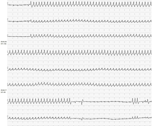
Ambulatory electrocardiography (ECG) monitoring revealed recurrent polymorphic ventricular tachycardia (PMVT). The arrhythmias persisted despite initial medical management, including calcium channel blockers and intravenous glyceryl trinitrate. The patient presented with recurrent palpitations and pre-syncope, with no chest pain.
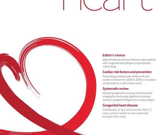
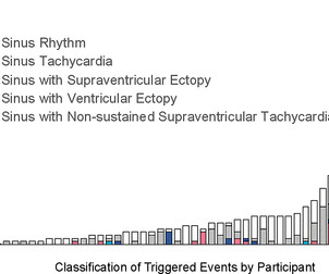
In adults with congenital heart disease (ACHD patients), atrial arrhythmias (AA) and heart failure (HF) are common. Early diagnosis and ECG documentation is therefore essential for arrhythmia management. Early diagnosis and ECG documentation is therefore essential for arrhythmia management.
Notwithstanding many insightful observations, the electrocardiogram (ECG) arguably ignited the big bang in our understanding of cardiac arrhythmias. Using ECG recording and deductive reasoning, our teachers and predecessors classified the bradycardias and tachycardias and proposed many mechanisms, subsequently proven to be correct.
We see here a wide complex tachycardia with a frequency of approx. The short VT after the end of the sustained ventricular tachycardia with the same QRS morphology also indicates a ventricular origin of this arrhythmia. 105-110 beats per minute that lasts for a good minute.
ECG#1 There is a regular tachycardia with a ventricular rate of about 180 bpm. Smith comment : When there is a regular wide complex tachycardia, first assess whether it is sinus or not. Put shortly is SVT with "Shark Fin STE" and not ventricular tachycardia. An ECG was recorded immediately and is shown below. Is there OMI?
A dditional M aterial on T oday's C ASE: — — Today’s E CG M edia P EARL # 37 ( 6:00 minutes Audio ) — Reviews how to determine IF Your Patient with an Arrhythmia is Hemodynamically Stable ! Figure: Following treatment with Atropine — the patient stabilized, and this 12-lead ECG was obtained.
Cingolani, director of Cardiogenetics and Preclinical Research in the Department of Cardiology in the Smidt Heart Institute at Cedars-Sinai, is exploring new ways to help patients with ventricular tachycardia (VT), a recurring, abnormally fast and irregular heartbeat that starts in the lower chambers, or ventricles, of the heart.
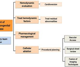
A multicenter study has described and validated a new strategy for guiding ablation procedures in patients with complex tachycardias. Ablation procedures use energy—usually heat or cold—to eliminate small areas of heart tissue that cause pathological cardiac arrhythmias, thereby restoring normal heart rhythm.
Sinus tachycardia – sinus rhythm above 100 bpm is a sinus tachycardia. Ventricular tachycardia – more than 7 consecutive complexes originating from ventricles at a rate of > 100 bpm. Supraventricular tachycardia – more than 7 consecutive complexes of supraventricular beats at a rate of > 100 bpm.
A prehospital 12-lead was recorded: There is a regular wide complex tachycardia. The computer diagnosed this as Ventricular Tachycardia. There is a wide complex regular tachycardia at a rate of 226. Toothache, incidental Wide Complex Tachycardia Could it be fascicular VT or Bundle Branch VT ( i.e., idiopathic VT )?
Background Cardiac magnetic resonance (CMR) allows comprehensive myocardial tissue characterisation, revealing areas of myocardial inflammation or fibrosis that may predispose to ventricular arrhythmias (VAs). A change in diagnosis after use of CMR ranged from 21% to 66% with a pooled average of 35% (29%–41%).
Sympathetic nervous system hyperactivity plays a major role in the pathogenesis of ventricular arrhythmias following myocardial infarction (MI).1,2 1,2 The stellate ganglia are an important nexus point for sympathetic innervation to the heart.3
She had a single chamber ICD/Pacemaker implanted several years prior due to ventricular tachycardia. Answer : The ECG above shows a regular wide complex tachycardia. Said differently, the ECG shows a rather slow ventricular tachycardia with a 2:1 VA conduction. Cardiac output (CO) was being maintained by the tachycardia.
(MedPage Today) -- Continuous rhythm monitoring showed the timeline in which heart rate and atrial tachycardias typically arise after binge drinking by young adults, researchers reported. In people with no known history of cardiac arrhythmias.
We reported the case of a 51-year-old woman who experienced multiple types of arrhythmias over three decades and was diagnosed with Danon disease late by genetic testing. Case summary A 51-year-old woman with a 36-year history of intermittent palpitations was admitted due to hemodynamically stable ventricular tachycardia (VT).
BackgroundAtrial fibrillation (AF) is the most common arrhythmia worldwide. The most common presenting complaints and ECG abnormality were trauma (44%) and sinus tachycardia (15%), respectively. Patients had routine 12-lead electrocardiograms (ECGs) regardless of presenting complaints.
Shortly after isoprenalin infusion was initiated, there were short runs of ventricular tachycardia. VT is the second most common presenting arrhythmia. Vaso or inotropic medications are not harmless, and can precipitate life threatening arrhythmias. She was started on isoprenalin (isoproterenol).
Ventricular tachycardia is a potentially life threatening cardiac arrhythmia. On the ECG, ventricular tachycardia can be defined as three or more ventricular ectopic beats occurring in a sequence at a rate more than 100 per minute. Another rare form of ventricular tachycardia is bidirectional ventricular tachycardia.
We see here a wide complex tachycardia with a frequency of approx. The short VT after the end of the sustained ventricular tachycardia with the same QRS morphology also indicates a ventricular origin of this arrhythmia. 105-110 beats per minute that lasts for a good minute.
Background Cardiac arrhythmias have been observed among patients hospitalised with acute COVID-19 infection, and palpitations remain a common symptom among the much larger outpatient population of COVID-19 survivors in the convalescent stage of the disease. Participants were instructed to trigger the monitor for palpitations.
Stereotactic arrhythmia radioablation (STAR) is used as a rescue treatment for refractory ventricular tachycardia (VT) following unsuccessful radiofrequency catheter ablation (RFCA). The mid-term outcomes of STAR, however, remain poorly known.
Surgical modifications and hemodynamic changes increase the susceptibility to arrhythmias, impacting morbidity and mortality rates, with arrhythmias being the leading cause of hospitalizations and sudden deaths. Macroreentrant atrial tachycardias, particularly cavotricuspid isthmus-dependent flutter, are frequently reported.
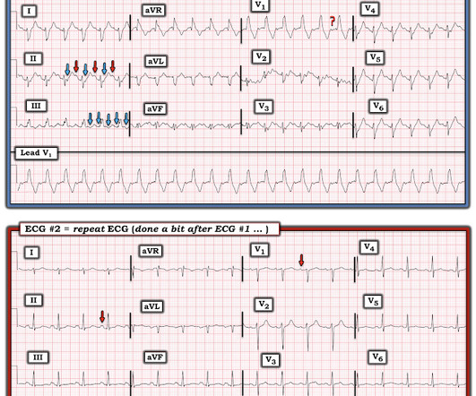
A series of cardiac arrhythmias were seen during the course of her resuscitation — including the interesting arrhythmia shown in the long lead II of Figure-1. PEARL # 5: The simple act of labeling P waves can be invaluable for solving an arrhythmia. Figure-6: Laddergram illustration of the mechanism in today's arrhythmia.
She was awake, alert, well perfused, with normal mental status and overall unremarkable physical exam except for a regular tachycardia, possible rales at both bases, some mild RUQ abdominal tenderness. Thus, I believe it is a regular, monomorphic, wide complex tachycardia. Or it could simply still be classic VT. What is the Diagnosis?
111.87), arrhythmias were detected in 9 (33%) patients. Two patients developed chronic sinus tachycardia at 4 and 16 months and were treated with Beta-blockers after eliminating all causes of sinus tachycardia. We did not identify significant risk factors for arrhythmias post-HT. On a median follow-up of 35.07
Even with tachycardia and a paced QRS duration of ~0.16 In this specific case, Left Bundle Branch (LBB) area pacing was pursued to achieve cardiac resynchronization. (J J Am Coll Cardiol. second I immediately knew there is no way this relative increase in QT duration ( compared to the R-R interval ) is going to be "normal".
MY Initial Thoughts: In my experience — all-too-many emergency providers fail to appreciate the potential contribution that a brief ( 1-to-2 line ) history may convey when interpreting arrhythmias. when the usual negative P wave deflection of sinus tachycardia is nowhere to be found in lead V1 )? What do YOU think?
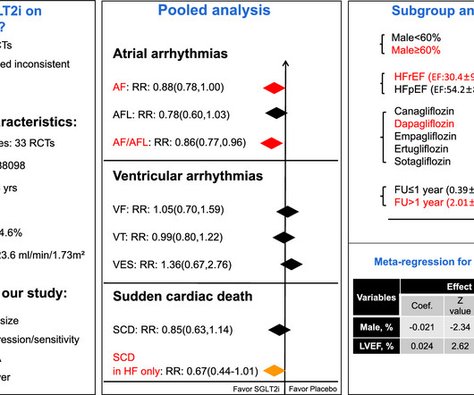
Objective We aimed to assess the effect of SGLT2i on arrhythmias by conducting a meta-analysis using data from randomized controlled trials(RCTs). Background Sodium-glucose co-transporter 2 inhibitors (SGLT2i) have shown cardioprotective effects via multiple mechanisms that may also contribute to decrease arrhythmias risk.
Stereotactic arrhythmia radioablation (STAR), used as a rescue treatment for refractory ventricular tachycardia (VT), still suffers from limitations to delineate the clinical target volume (CTV).
Catheter ablation as a treatment for ventricular tachycardia has lagged behind ablation procedures for atrial arrhythmias in becoming an established first-line therapy.
What is the most likely cause of this arrhythmia? IMPRESSION: Given the presence of a wide tachycardia — with 2 distinct QRS morphologies, and no sign of P waves — a presumed diagnosis of B i D irectional Ventricular Tachycardia has to be made. He developed cardiac arrest shortly after the ECG in Figure-1 was recorded.
Stereotactic arrhythmia radioablation (STAR) is a new modality for treating refractory ventricular tachycardia (VT) after failed catheter ablation (CA). Artificial intelligence (AI) may help predict the need for redo CA after STAR, however, predictive features that contribute most to the need for redo CA remain elusive.
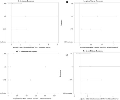
Objectives Examine the association between arrhythmias and adverse maternal outcomes in women with structurally normal hearts. Arrhythmia was previously diagnosed in 58.0% After adjusting for age, parity and comorbidities, the presence of any arrhythmia was an independent predictor of CS (OR 1.7 SVT cases but only in 9.7%
Background Computer-assisted interpretation of single-lead ECG is the preliminary method for clinicians to flag and further evaluate an arrhythmia of clinical importance for acutely ill patients. We calculated classifier statistics for each arrhythmia, all arrhythmias and strips where the model identified normal sinus rhythm.
The only exceptions that I'm aware of to the above-cited morphologic criteria for VT are: i ) IF the rhythm is antidromic AVRT in which case the impulse travels forward over an AP ( A ccessory P athway ) in a patient with WPW, therefore resulting in a regular WCT rhythm that resembles VT ( For more on the various arrhythmias in patients with WPW (..)
Note the Timed Contents that I detail below facilitate finding specific material. == ECG Podcast #4 — All About Comparison ECGs for 12-Leads and Arrhythmias ( Comparing ECGs seems so "easy" to do — but so often is not done correctly! ) — published by Mayo Clinic CV Podcast Series on 5/21/2024 ( 35 minutes ). What are the problems?
BackgroundHuman pluripotent stem cell-derived cardiomyocytes (hPSC-CMs) show tremendous promise for cardiac regeneration following myocardial infarction (MI), but their transplantation gives rise to transient ventricular tachycardia (VT) in large-animal MI models, representing a major hurdle to translation.
Although not immediately apparent ( because it is hard to distinguish the limits of a P wave in lead II ) — the rhythm is Sinus Tachycardia , as other leads correspond to the timing of what seems to be an upright deflection that is subtly notching the end of the T wave in lead II ( and which almost certainly represents the sinus P wave ).
A 21-year-old woman presented to arrhythmia clinic with episodes of recurrent paroxysmal rapid palpitations. We found a narrow complex tachycardia of cycle 460 ms length ms which was repeatedly induced with atrial extrastimuli. What is the mechanism of the tachycardia?
We organize all of the trending information in your field so you don't have to. Join thousands of users and stay up to date on the latest articles your peers are reading.
You know about us, now we want to get to know you!
Let's personalize your content
Let's get even more personalized
We recognize your account from another site in our network, please click 'Send Email' below to continue with verifying your account and setting a password.


Let's personalize your content