This site uses cookies to improve your experience. To help us insure we adhere to various privacy regulations, please select your country/region of residence. If you do not select a country, we will assume you are from the United States. Select your Cookie Settings or view our Privacy Policy and Terms of Use.
Cookie Settings
Cookies and similar technologies are used on this website for proper function of the website, for tracking performance analytics and for marketing purposes. We and some of our third-party providers may use cookie data for various purposes. Please review the cookie settings below and choose your preference.
Used for the proper function of the website
Used for monitoring website traffic and interactions
Cookie Settings
Cookies and similar technologies are used on this website for proper function of the website, for tracking performance analytics and for marketing purposes. We and some of our third-party providers may use cookie data for various purposes. Please review the cookie settings below and choose your preference.
Strictly Necessary: Used for the proper function of the website
Performance/Analytics: Used for monitoring website traffic and interactions
Specific cardiovascular diseases, such as acute myocardial infarction, arrhythmias, pulmonary hypertension and pericarditis, were also pointed. Elevated risk of arrhythmias, particularly atrial fibrillation, correlated with occupational silica exposure.
a global leader in cardiac arrhythmia treatment and part of Johnson & Johnson MedTec h i , announced the submission of the VARIPULSE Platform for Premarket Approval Application (PMA) to the U.S. milla1cf Thu, 03/28/2024 - 07:28 March 28, 2024 — Biosense Webster, Inc., Food & Drug Administration ( FDA ).
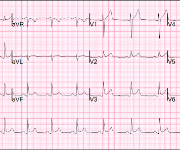
PR depression, which suggests pericarditis 4. We also showed that, of 47 cases of pericarditis with ST elevation, none had ST depression in aVL. ) I’ll add the following 2 comments: i ) This patient presumably has effusive-constrictive pericarditis. Absence of any ST depression in aVL. ( Clin Cardiol 22:334-344, 1999 ).
Circulation: Arrhythmia and Electrophysiology, Ahead of Print. Freedom from atrial arrhythmia was evaluated continuously through 12 months using standard rhythm monitoring for symptomatic episodes and 24-hour Holter at 6 and 12 months.RESULTS:Eighty-two patients (74% male, 51.2% paroxysmal, and 58.5% deep sedation) were treated.
Pericarditis? If you were thinking that this is pericarditis, that would be possible in the absence of any clinical information. However, there is zero PR depression which would be VERY unusual in pericarditis. P.S.: Acute pericarditis may produce diffuse ST elevation. Presence of STD is helpful; absence is not.
Circulation: Arrhythmia and Electrophysiology, Ahead of Print. Colchicine did not prevent atrial arrhythmia recurrence at 2 weeks (31% versus 32%; hazard ratio [HR], 0.98 [95% CI, 0.59–1.61];P=0.92) Postablation chest pain consistent with pericarditis was reduced with colchicine (4% versus 15%; HR, 0.26 [95% CI, 0.09–0.77];P=0.02)
Haven't you been taught that this favors pericarditis? Weren't you taught that concave morphology favors pericarditis? Expert ECG interpretation can often distinguish normal variant STE from OMI from pericarditis. There is now a regular sinus rhythm ( there had previously been a fairly marked sinus arrhythmia in ECG #1 ).
ECG of pneumopericardium and probable myocardial contusion shows typical pericarditis Male in 30's, 2 days after Motor Vehicle Collsion, complains of Chest Pain and Dyspnea Head On Motor Vehicle Collision. Other Arrhythmias ( PACs, PVCs, AFib, Bradycardia and AV conduction disorders — potentially lethal VT/VFib ). ST depression.
T-wave alternans and the susceptibility to ventricular arrhythmias. Chronic amiodarone evokes no torsade de pointes arrhythmias despite QT lengthening in an animal model of acquired long-QT syndrome. Both ST segment and T wave alternans have been known to precede malignant ventricular arrhythmias. Pacing Clin Electrophysiol.
Cardiac involvement in multisystemic inflammatory syndrome in adults has been described mainly in young men without other comorbidities and may present with different clinical scenarios, including acute heart failure, life‐threatening arrhythmias, pericarditis, and myocarditis, with a nonnegligible risk of mortality (up to 7% of all cases).
Realizing that slight variation in the P-P interval is common ( known as sinus arrhythmia ) — the PINK arrows in Figure-3 suggest the probable location of underlying sinus P waves. Certain complex arrhythmias may have more than a single plausible rhythm interpretation. This is precisely what we see in Figure-4.
This is the proposed mechanism of precipitation of arrhythmias in Brugada syndrome during febrile episodes. There is a potential risk for drug challenge in that life threatening ventricular arrhythmias could be precipitated. This leads to shortening of action potential duration. With proper precautions, risk can be reduced.
He has a family history concerning for arrhythmia. Given the circumstances of his car crash, we presume it was due to an underlying arrhythmia. He has a family history concerning for arrhythmia with his father requiring some sort of device (PPM, ICD, unclear) at a young age. ST depression. Myocardial Contusion?
The rhythm is uncertain ( ie, We only see 4 beats — because the same 4 beats are repeated in limb and chest leads — but in lead II there appears to be sinus bradycardia and arrhythmia plus a P wave with a PR interval too short to conduct preceding beat #1 — therefore need for a longer period of monitoring ).
The "flu-like" illness suggests myo- or pericarditis, but that would be a diagnosis of exclusion. Jenkins and Frick — I offer 3 additional examples of artifactual distortion ( excerpted from my ECG Blog ) — that resulted in arrhythmia misdiagnosis. Do not wait for the troponin; a lot of myocardium will be dead if you do.
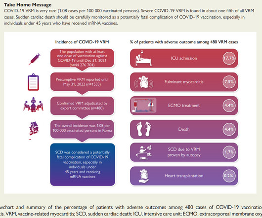
The KDCA also established a reporting system with a legal obligation for special adverse events including myocarditis and pericarditis after COVID-19 vaccination. It’s pretty clear now that the descriptors used for the vaccine for much of the pandemic were hyperbolic and lacked nuance.
Measurement of QT interval is very important, as QT prolongation can predispose to serious, life threatening ventricular arrhythmias. QT prolongation leads to torsades des pointes, which is a very serious arrhythmia. PR segment elevation and depression can occur in atrial infarction and pericarditis. to 0.43, normal range.
The second most common cause of medical cardiac tamponade is acute idiopathic pericarditis. Less common etiologies include uremia, bacterial or tubercular pericarditis, chronic idiopathic pericarditis, hemorrhage, and other causes such as autoimmune diseases, radiation, myxedema, etc.
First Troponin I was <2 and peak was 8, echo showed subtle apical lateral hypokinesis, CRP was elevated, and patient was discharged with a diagnosis of regional pericarditis. In this case, there would be evolution, but the evolution would be typical of pericarditis (if the diagnosis of pericarditis was accurate!!
In August, the CDC reported 29 cases of pericarditis, including five in persons with a history of pericarditis after mRNA COVID-19 vaccine ; 10 Importantly, the Novovax vaccine is a protein based vaccine that was hoped to not be associated with myocarditis as was noted with the mRNA vaccines.
Though less prevalent in younger patients, occlusion MI may occur and requires the same early interventions as older patients. - - Pericarditis and myocarditis should be a diagnosis of exclusion. My Thoughts on the ECG in Figure-1: The rhythm in ECG #1 is sinus arrhythmia. Figure-1: I've labeled the initial ECG in today's case.
While traditionally described as “benign early repolarization”, they have been associated with J wave syndromes along with Brugada syndrome, causing ventricular arrhythmias (1, 2). Prominent J waves and ventricular fibrillation caused by myocarditis and pericarditis after BNT162b2 mRNA COVID-19 vaccination. J wave syndromes.
Overnight telemetry showed no arrhythmias ( important to reduce the risk of worrisome arrhythmia given this patient's chief complaint of sudden syncope without prodrome ). Smith : I recognize this as a STEMI mimic. I was not alarmed. The providers showed me the ECG and I told them that I thought it was a fake. Troponins were negative.
We organize all of the trending information in your field so you don't have to. Join thousands of users and stay up to date on the latest articles your peers are reading.
You know about us, now we want to get to know you!
Let's personalize your content
Let's get even more personalized
We recognize your account from another site in our network, please click 'Send Email' below to continue with verifying your account and setting a password.


Let's personalize your content