This site uses cookies to improve your experience. To help us insure we adhere to various privacy regulations, please select your country/region of residence. If you do not select a country, we will assume you are from the United States. Select your Cookie Settings or view our Privacy Policy and Terms of Use.
Cookie Settings
Cookies and similar technologies are used on this website for proper function of the website, for tracking performance analytics and for marketing purposes. We and some of our third-party providers may use cookie data for various purposes. Please review the cookie settings below and choose your preference.
Used for the proper function of the website
Used for monitoring website traffic and interactions
Cookie Settings
Cookies and similar technologies are used on this website for proper function of the website, for tracking performance analytics and for marketing purposes. We and some of our third-party providers may use cookie data for various purposes. Please review the cookie settings below and choose your preference.
Strictly Necessary: Used for the proper function of the website
Performance/Analytics: Used for monitoring website traffic and interactions
Autonomic dysfunction caused by spinal cord injury is associated with abnormalities in blood pressure, heart rate variability, arrhythmias and blunted cardiovascular response to exercise which can limit the capacity to perform physical activity [1]. Am J Phys Med Rehabil. 2007 Feb;86(2):142-52. doi: 10.1097/PHM.0b013e31802f0247. 2023.12.010.
Cerebrovascular damage can cause cardiac arrhythmias related to disinhibition of right insular cortex with resulting increased sympathetic tone. ECG changes resembling ST elevation myocardialinfarction has also been described after traumatic intracranial hemorrhage [4]. In a study of 204 subjects, 31% had troponin elevation.
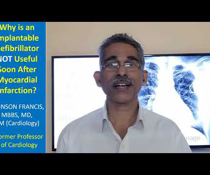
Ventricular tachycardia is a potentially life threatening cardiac arrhythmia. Those at risk of recurrent VT with previous myocardialinfarction and left ventricular dysfunction also need an implantable defibrillator. If the rate is very fast, hemodynamic deterioration can occur rapidly.
Angina is another common symptom due the hypertrophy which causes a coronary supply demand mismatch Symptoms of HCM include syncope/near syncope, which could be precipitated by exertion, hypovolemia, rapid standing, Valsalva manoeuvre, diuretics, vasodilators or arrhythmia. Palpitations can be felt if there are arrhythmias.
Measurement of QT interval is very important, as QT prolongation can predispose to serious, life threatening ventricular arrhythmias. QT prolongation leads to torsades des pointes, which is a very serious arrhythmia. This represents the ventricular depolarization as well as repolarization. One easy value which I remember is 0.34
It can automatically detect life threatening ventricular arrhythmias and treat them, either with a shock or, sometimes by overdrive pacing. You may be knowing that one of the causes for inappropriate ICD shocks, is supraventricular arrhythmia, wrongly detected by the defibrillator as a ventricular arrhythmia and giving a shock.
So this is a typical Brugada syndrome ECG, which can be easily mistaken for an acute myocardialinfarction with ST elevation in anterior leads may be taken as STEMI if the person presents with chest pain for some other reason. An interesting fact is that many of the persons experience arrhythmias in Brugada syndrome with fever.
We organize all of the trending information in your field so you don't have to. Join thousands of users and stay up to date on the latest articles your peers are reading.
You know about us, now we want to get to know you!
Let's personalize your content
Let's get even more personalized
We recognize your account from another site in our network, please click 'Send Email' below to continue with verifying your account and setting a password.


Let's personalize your content