This site uses cookies to improve your experience. To help us insure we adhere to various privacy regulations, please select your country/region of residence. If you do not select a country, we will assume you are from the United States. Select your Cookie Settings or view our Privacy Policy and Terms of Use.
Cookie Settings
Cookies and similar technologies are used on this website for proper function of the website, for tracking performance analytics and for marketing purposes. We and some of our third-party providers may use cookie data for various purposes. Please review the cookie settings below and choose your preference.
Used for the proper function of the website
Used for monitoring website traffic and interactions
Cookie Settings
Cookies and similar technologies are used on this website for proper function of the website, for tracking performance analytics and for marketing purposes. We and some of our third-party providers may use cookie data for various purposes. Please review the cookie settings below and choose your preference.
Strictly Necessary: Used for the proper function of the website
Performance/Analytics: Used for monitoring website traffic and interactions
Notwithstanding many insightful observations, the electrocardiogram (ECG) arguably ignited the big bang in our understanding of cardiac arrhythmias. The contemporaneous evolution of cardiac surgery permitted direct access to the heart to modify arrhythmia substrate.
The data was presented by Vivek Reddy , MD, Director of Cardiac Arrhythmia Services at The Mount Sinai Hospital , during the European Heart Rhythm Association (EHRA) conference in Berlin, Germany. milla1cf Fri, 04/12/2024 - 20:53 April 12, 2024 — HeartBeam, Inc. , The HeartBeam technology gathers far more data than a single-lead ECG.
The treatment of outflow tract ventricular arrhythmias (OTVA) through radiofrequency ablation requires the precise identification of the site of origin (SOO). Current clinical methods to identify the SOO are based on qualitative analysis of pre-operative electrocardiograms (ECG), heavily relying on physician’s expertise.
In a world where technology reigns supreme, one of the most profound tools in medicine remains the irreplaceable electrocardiogram (ECG). An abnormal electrocardiogram can mean many things. Other times, an irregular recording can signal a medical emergency, such as a myocardial infarction or a dangerous arrhythmia.
BackgroundAtrial fibrillation (AF) is the most common arrhythmia worldwide. Patients had routine 12-lead electrocardiograms (ECGs) regardless of presenting complaints.
Persistent cardiac arrhythmias are readily amenable to detection by performing a standard electrocardiogram (ECG), but detection of transient (paroxysmal) arrhythmias has long been a significant cause of frustration to both doctors and patients.
Accurate diagnosis of arrhythmia is crucial for timely and effective treatment. Electrocardiogram (ECG) plays a key role in the diagnosis of arrhythmia. This provides a new idea for ECG based arrhythmia diagnosis.
Thirty day electrocardiogram (ECG) monitoring in patients with hypertrophic cardiomyopathy (HCM) detects more arrhythmias than the standard 24 to 48 hours, according to late breaking science presented at EHRA 2023, a scientific congress of the European Society of Cardiology (ESC).
A gradual increase in arrhythmia recurrences during 12 months after catheter ablation (CA) of atrial fibrillation (AF) is still reported.1 A gradual increase in arrhythmia recurrences during 12 months after catheter ablation (CA) of atrial fibrillation (AF) is still reported.1
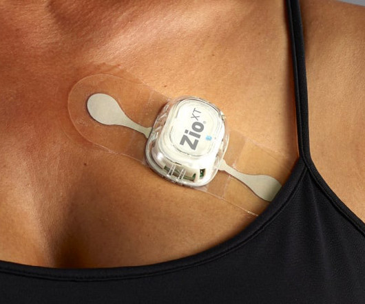
The Zio monitor ECG System secured its CE mark based on compliance to EU MDR standards of performance, quality, safety, and efficacy, along with the body of clinical evidence supporting Zio in detecting potential cardiac arrhythmias. Diagnostic Utility of a Novel Leadless Arrhythmia Monitoring Device, American Journal of Cardiology , 2013.
Familial ST-depression Syndrome (Fam-STD) is a recently identified inherited cardiac disease characterized by a distinct electrocardiographic phenotype and occurrence of arrhythmias and heart failure.
The experimental results show that the proposed technique is superior to the existing wavelet based approach and NLM filtering, with the higher SNR and structure similarity index measure (SSIM), the lower root mean squared error (RMSE) and percent root mean square difference (PRD).ConclusionsThe
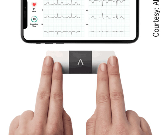
The Kardia 12L ECG System, featuring a game-changing patented technology, is the world’s first AI-powered handheld 12-lead electrocardiogram ( ECG ) system with a unique single-cable design. KAI 12L employs multiple deep neural network algorithms, trained and validated on more than 1.75 million ECGs from leading US medical centers.
Electrocardiogram (ECG) is a simple, noninvasive screening method for cardiovascular disease and arrhythmia. The impact of ECG abnormality on mortality is not certain in low-risk populations.
Although several studies have reported on estimating the risk of developing fatal arrhythmias from ECG findings, the use of ECG to identify the severity of heart failure (HF) by applying deep learning (DL) methods has not been established. Objectives In hypertrophic cardiomyopathy (HCM), specific ECG abnormalities are observed.
Diagnosis and assessment of disease progression in arrhythmogenic cardiomyopathy (ACM) are currently based on the 2010 Task Force Criteria (TFC) with the 12-lead electrocardiogram (ECG) being an important modality. The presence of inverted T-waves might be associated with lethal ventricular arrhythmias.
Patients with documented spontaneous type-1 (Spont-Type-I) Brugada syndrome (BrS) electrocardiogram (ECG) are at higher risk of arrhythmias compared to those with a drug-induced type I (DI-Type-I) ECG (1).
Electrocardiogram in clinic showed sinus arrhythmia with early repolarization and no ischemic changes. A 14-year-old male with no significant medical history presented with intermittent palpitations for 2–3 months that occurred at rest and were associated with light-headedness.
Background Studies in small animals and human patients have suggested that anthracyclines may prolong cardiac repolarization, or at least inhibit repolarization reserve, predisposing to QT prolongation and dangerous arrhythmias such as Torsades de pointes.
Circulation: Arrhythmia and Electrophysiology, Ahead of Print. While any arrhythmia patient may recur after acutely successful ablation, AF is unusual in that patients may have long-term arrhythmia freedom despite a lack of acute success.
Cerebrovascular damage can cause cardiac arrhythmias related to disinhibition of right insular cortex with resulting increased sympathetic tone. Lead electrocardiogram changes after supratentorial intracerebral hemorrhage. Lead electrocardiogram changes after supratentorial intracerebral hemorrhage. PMID: 7355693.
QT prolongation and the occurrence of ventricular arrhythmias with exercise are another important aspect of exercise testing in children. Ventricular arrhythmias during exercise can be documented in congenital long QT syndromes as well as in catecholaminergic polymorphic ventricular tachycardia.
A 12-lead electrocardiogram revealed a narrow QRS complex tachycardia with a rate of 157 beats per minute and a prolonged RP relationship. Post-procedure, the patient remained free from arrhythmia and showed restored normal cardiac function and was prescribed a low-dose -blocker for 1 month.
Methods The study enrolled patients with frequent PVCs who were not combined with other arrhythmias or structural heart disease. The demographic, clinical, and electrocardiogram features of the patients in each group were compared. This study aims to analyze the palpitations and related factors in patients with frequent PVCs.
Preimplant electrocardiogram screening is recommended to prevent implantation in patients at high risk of T wave over-sensing. The S-ICD is implanted subcutaneously or intramuscularly with the generator placed in the left midaxillary line and the lead tunneled subcutaneously in the left para-sternal region.
Introduction Electrocardiogram (ECG) machines are vital in diagnosing and monitoring various cardiac conditions. Understanding ECG Machines Electrocardiogram machines are medical devices used to record the heart’s electrical activity.
Follow-up contained regular visits at our outpatient clinic at 1, 3, 6, and 12 months including 7-day Holter electrocardiograms. After a blanking period of 6 weeks, recurrence of any atrial arrhythmia was documented in 26 patients (52%). Most arrhythmia recurrences were reentrant AT. to 202.2 ± 21.6 ms After a total of 1.46 ± 0.68
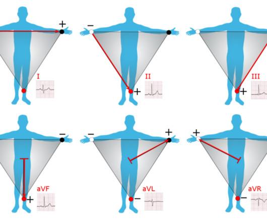
Introduction to ECG Testing & Einthoven’s Triangle: The electrocardiogram (ECG) represents the heart’s electrical activity, resulting from the contraction (depolarization) and relaxation (repolarization) of the atrial and ventricular muscles. Let’s start with grasping the basics of electrocardiography.
A Holter monitor is a portable electrocardiogram (ECG) device. A major benefit of the long-term constant monitoring provided by these devices is that they can help diagnose arrhythmias that other tests, like an EKG, cannot detect. What is a Holter monitor ? What are the main differences between Holter monitors vs. loop recorders?
Electrocardiograms (EKG or ECG): This diagnostic method effectively detects irregular heartbeats or arrhythmias by analyzing the electrical signals in the heart. Recommended tests, including routine lab work, blood pressure checks, and other tests covered below, are vital to assess heart health.
A deep neural network for 12-lead electrocardiogram interpretation outperforms a conventional algorithm, and its physician over-read, in the diagnosis of atrial fibrillation. How can you avoid overlooking this arrhythmia? The reasons for overlooking this arrhythmia are simple: True MAT is not a common rhythm. Poon et al.
The attending provider wrote “Agree with electrocardiogram interpretation”. At this point, there was no improvement in LV function and he was out of the convalescent phase of his MI, so the decision was made to install an ICD for arrhythmia prophylaxis. The computer diagnostic algorithm diagnosed “Sinus rhythm. Normal EKG”. Normal ECG.
Traditional tools like stethoscopes, blood pressure gauges, and electrocardiograms (ECG) are fundamental for standard diagnostic practices. Implantable Devices Implantable cardiac devices, such as pacemakers and implantable cardioverter-defibrillators (ICDs), are crucial in managing arrhythmias and other cardiac conditions.
New insights into the use of the 12-lead electrocardiogram for diagnosing acute myocardial infarction in the emergency department. All electrocardiograms (ECGs) and coronary angiograms were blindly analyzed by experienced cardiologists. Harhash AA, Huang JJ, Reddy S, et al. aVR ST segment elevation: acute STEMI or not?
The 2 clinical areas of most potential benefit to emergency care from computerized interpretations are: i ) Cardiac arrhythmias; and , ii ) Rapid detection of acute coronary O cclusion ( ie, detection of acute O MI ) in cases for which easily recognizable STEMI-criteria are not present. J Am Bd Fam Prac 1:17-24, 1989.
In a study published in Communications Medicine , David Ouyang, MD, assistant professor of Cardiology and Medicine at Cedars-Sinai, along with Chugh and fellow investigators trained a deep learning algorithm to study patterns in electrocardiograms, also known as ECGs, which are recordings of the heart’s electrical activity.
You will note that it is essentially an unremarkable electrocardiogram except for some PACS. No arrhythmias occurred en route. At baseline, the patient has some expected, normal STE in lead V2, further demonstrating that the STD morphology in the presentation ECG above is "true" and diagnostic.
Electrocardiogram (ECG) abnormalities can be found in almost all patients, with Wolff–Parkinson–White (WPW) syndrome being the most common. We reported the case of a 51-year-old woman who experienced multiple types of arrhythmias over three decades and was diagnosed with Danon disease late by genetic testing.
In this setting, family history of arrhythmia and being carrier of a pathogenic/likely pathogenic variant are the main risk factors for LV systolic dysfunction, while LV global longitudinal strain (LV-GLS) and Holter electrocardiogram (ECG) showed a relevant role in terms of prediction of LV systolic dysfunction and outcomes.
Arrhythmias (Irregular Heartbeats) Persistent abnormal heart rhythms can disrupt the heart’s pumping efficiency, eventually causing it to enlarge to maintain blood flow. Electrocardiogram (ECG/EKG) An ECG records the electrical activity of the heart and can help detect abnormalities in the heart’s rhythm that might contribute to enlargement.
Examination of previously reported predictors of lethal ventricular arrhythmias or sudden cardiac death (VA/SCD) by ECG in Brugada syndrome (BrS) is a manual measurement and lacks objectivity.
Recognition of distinct arrhythmia syndromes over the last 3 decades, such as long QT syndrome, Brugada syndrome, catecholaminergic polymorphic ventricular tachycardia, and early repolarization (ER) syndrome (ERS), has changed this field, and the diagnosis of IVF has substantially decreased.1
We organize all of the trending information in your field so you don't have to. Join thousands of users and stay up to date on the latest articles your peers are reading.
You know about us, now we want to get to know you!
Let's personalize your content
Let's get even more personalized
We recognize your account from another site in our network, please click 'Send Email' below to continue with verifying your account and setting a password.


Let's personalize your content