This site uses cookies to improve your experience. To help us insure we adhere to various privacy regulations, please select your country/region of residence. If you do not select a country, we will assume you are from the United States. Select your Cookie Settings or view our Privacy Policy and Terms of Use.
Cookie Settings
Cookies and similar technologies are used on this website for proper function of the website, for tracking performance analytics and for marketing purposes. We and some of our third-party providers may use cookie data for various purposes. Please review the cookie settings below and choose your preference.
Used for the proper function of the website
Used for monitoring website traffic and interactions
Cookie Settings
Cookies and similar technologies are used on this website for proper function of the website, for tracking performance analytics and for marketing purposes. We and some of our third-party providers may use cookie data for various purposes. Please review the cookie settings below and choose your preference.
Strictly Necessary: Used for the proper function of the website
Performance/Analytics: Used for monitoring website traffic and interactions
Arrhythmias (Irregular Heartbeats) Persistent abnormal heart rhythms can disrupt the heart’s pumping efficiency, eventually causing it to enlarge to maintain blood flow. Electrocardiogram (ECG/EKG) An ECG records the electrical activity of the heart and can help detect abnormalities in the heart’s rhythm that might contribute to enlargement.
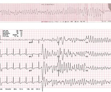
During the intravenous lacosamide infusion, the patient developed sudden cardiac arrest caused by ventricular arrhythmias necessitating resuscitation. Workup including routine laboratory results, 12-lead electrocardiogram (ECG), echocardiogram, and coronary angiogram was non-specific.
It is also published in Heart Rhythm , the official journal of the HRS, Journal of Arrhythmia , the official journal of the APHRS, and Journal of Interventional Cardiac Electrophysiology , the official journal of the LAHRS. Antiarrhythmic drugs are advised for some patients to prevent arrhythmia recurrences early after the procedure.
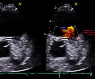
Electrocardiogram in clinic showed sinus arrhythmia with early repolarization and no ischemic changes. The echocardiogram showed normal cardiac structure and function, however, there was a concern for possible anomalous origin of the left coronary artery.
However, an echocardiogram is a different test, also conducted for heart activity. An electrocardiogram is a machine used to record the heart's electrical activity. Arrhythmia In simple words, arrhythmia refers to an irregular heartbeat. ECG and EKG refer to the same thing.
Yes, COVID-19 symptoms can resemble a heart attack, including chest pain, shortness of breath, and changes in echocardiogram or EKG. Electrocardiograms (EKG or ECG): This diagnostic method effectively detects irregular heartbeats or arrhythmias by analyzing the electrical signals in the heart.
Echocardiogram is indicated (Correct) C. Start aspirin and Plavix Correct answer: (B) (B) Echocardiogram is indicated. Explanation: Shown electrocardiogram suggests left ventricular hypertrophy. Shown electrocardiogram suggests left ventricular hypertrophy. No further workup is indicated B. Start with a Free Trial.
The attending provider wrote “Agree with electrocardiogram interpretation”. An echocardiogram showed severely reduced global systolic function with an EF of 20-25% and an LV apical thrombus. An echocardiogram showed an EF of 20-25%. The computer diagnostic algorithm diagnosed “Sinus rhythm. Normal EKG”. Normal ECG.
Formal echocardiogram showed normal EF, no wall motion abnormalities, no pericardial effusion. Prior to Mizusawa's study, it was thought that the incidence of syncope, arrhythmia, or SCD in this cohort was low [7]. Induced Brugada-type electrocardiogram, a sign for imminent malignant arrhythmias. There was a 0.9%
Seventh , an immediate echocardiogram can make the distinction. Sixth , placement of posterior leads (take leads V4-V6 and place them at the level of the tip of the scapula, with V4 placed at the posterior axillary line ("V7"), V6 at paraspinal area ("V9"), and V5 ("V8") between them. At lease 0.5 Kligfield P, Gettes LS, Bailey JJ, et al.
Traditional tools like stethoscopes, blood pressure gauges, and electrocardiograms (ECG) are fundamental for standard diagnostic practices. This transformation extends to the use of machine learning (ML) algorithms developed by startups, which analyze medical imaging data such as ECGs, echocardiograms, and cardiac MRI scans.
A formal echocardiogram was completed the next day and again showed a normal ejection fraction without any focal wall motion abnormalities to suggest CAD. Prior to Mizusawa's study, it was thought that the incidence of syncope, arrhythmia, or SCD in this cohort was low [7]. There was a 0.9% per year incidence of SCD in this cohort [1].
See this case: what do you think the echocardiogram shows in this case? New insights into the use of the 12-lead electrocardiogram for diagnosing acute myocardial infarction in the emergency department. All electrocardiograms (ECGs) and coronary angiograms were blindly analyzed by experienced cardiologists.
You will note that it is essentially an unremarkable electrocardiogram except for some PACS. No arrhythmias occurred en route. Unfortunately there is no echocardiogram accessible because the patient checked himself out of the hospital in order to get back to his home state before it could be completed.
Abnormal Electrocardiogram (ECG): Defined (San Fran syncope rule) as any new changes when compared to the last ECG or presence of non-sinus rhythm. Finally, much of this correlates well with The new Canadian Syncope Arrhythmia Risk Score , just published in 2016, results of which are given below in the Annotated Bibliography.
His cardiac testing completed to date consist of an electrocardiogram and an echocardiogram performed Feb 16th, 2023 that were both normal. The pain resolved a few weeks later. He has had COVID twice, first in September of 2020, and his second time in January of 2023.
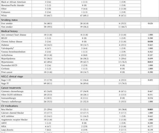
Retrospective analysis of NSCLC patients with 1 echocardiogram post-osimertinib between 2007 and 2022 was performed. There was an increased risk of grade 2 cardiac CTCAE with pre-existing arrhythmia [hazard ratio(HR) 3.90, 95%CI, 1.1113.72; p=0.034] and higher body mass index (HR 1.07, 95%CI, 1.001.14; p=0.04). and 8.5%, respectively.
We organize all of the trending information in your field so you don't have to. Join thousands of users and stay up to date on the latest articles your peers are reading.
You know about us, now we want to get to know you!
Let's personalize your content
Let's get even more personalized
We recognize your account from another site in our network, please click 'Send Email' below to continue with verifying your account and setting a password.


Let's personalize your content