This site uses cookies to improve your experience. To help us insure we adhere to various privacy regulations, please select your country/region of residence. If you do not select a country, we will assume you are from the United States. Select your Cookie Settings or view our Privacy Policy and Terms of Use.
Cookie Settings
Cookies and similar technologies are used on this website for proper function of the website, for tracking performance analytics and for marketing purposes. We and some of our third-party providers may use cookie data for various purposes. Please review the cookie settings below and choose your preference.
Used for the proper function of the website
Used for monitoring website traffic and interactions
Cookie Settings
Cookies and similar technologies are used on this website for proper function of the website, for tracking performance analytics and for marketing purposes. We and some of our third-party providers may use cookie data for various purposes. Please review the cookie settings below and choose your preference.
Strictly Necessary: Used for the proper function of the website
Performance/Analytics: Used for monitoring website traffic and interactions
He was defibrillated into VT. He then underwent dual sequential defibrillation into asystole. The ECG shows severe ischemia, possibly posterior OMI. But cardiac arrest is a period of near zero flow in the coronary arteries and causes SEVERE ischemia. It takes time for that ischemia to resolve. They started CPR.
It should be kept in mind that on occasions, beta-one agonist can result in increased ventricular ectopy e.g., in severe myocardial ischemia (by increasing myocardial demand), or sometimes with congenital long-QT syndrome. Smith, this can be accomplished by either using beta-one agonists or temporary transvenous pacing.
Ventricular tachycardia is a potentially life threatening cardiac arrhythmia. Monomorphic ventricular tachycardia in the setting of acute myocardial ischemia can also be treated by intravenous lignocaine bolus followed by infusion. If the rate is very fast, hemodynamic deterioration can occur rapidly.
She was successfully revived after several rounds of ACLS including defibrillation and amiodarone. T-wave alternans and the susceptibility to ventricular arrhythmias. Chronic amiodarone evokes no torsade de pointes arrhythmias despite QT lengthening in an animal model of acquired long-QT syndrome. Pacing Clin Electrophysiol.
She underwent cardiopulmonary resuscitation for VT/VFib — with ROSC ( R eturn O f S pontaneous C irculation ) following defibrillation and treatment with Epinephrine and Amiodarone. A series of cardiac arrhythmias were seen during the course of her resuscitation — including the interesting arrhythmia shown in the long lead II of Figure-1.
We examined the effect of ibutilide, a class III antiarrhythmic agent, on the energy requirement for atrial defibrillation and assessed the value of this agent in facilitating cardioversion in patients with atrial fibrillation that is resistant to conventional transthoracic cardioversion. 25, 2022 ).
She was never defibrillated. Therefore, she underwent temporary pacemaker placement and overdrive pacing at a rate of 90 bpm to keep the heart rate up in order to prevent these PVCs triggering ventricular arrhythmia. Hypokalemia was unlikely because she continued to have ventricular arrhythmia despite of correcting electrolytes.
12 minutes later, the patient went back into VFib arrest and underwent another 15 minutes of resuscitation followed by successful defibrillation and sustained ROSC. In total, he received approximately 40 minutes of CPR and 7 defibrillation attempts. An ICD was placed due to suspicion of a primary arrhythmia.
However, he suddenly developed a series of malignant ventricular arrhythmias. He required multiple defibrillations within a period of a few hours. Below are printouts of some of the arrhythmias recorded. There is no definite evidence of acute ischemia. (ie, What do you think?
What is the most likely cause of this arrhythmia? Acute myocardial ischemia. Despite prolonged resuscitation with multiple defibrillation attempts — the patient could not be saved. = He developed cardiac arrest shortly after the ECG in Figure-1 was recorded. QUESTIONS: How would YOU interpret the ECG in Figure-1 ?
This may result in ischemia (lack of oxygen to the heart muscle), causing parts of the heart to weaken and enlarge. Arrhythmias (Irregular Heartbeats) Persistent abnormal heart rhythms can disrupt the heart’s pumping efficiency, eventually causing it to enlarge to maintain blood flow.
The first task when assessing a wide complex QRS for ischemia is to identify the end of the QRS. The ST segment changes are compatible with severe subendocardial ischemia which can be caused by type I MI from ACS or potentially from type II MI (non-obstructive coronary artery disease with supply/demand mismatch). What do you think?
V1 sits over both the RV and the septum, so transmural ischemia of either one with give OMI pattern in V1 and reciprocal STD in V5 and V6. Case progression: The automated EKG interpretation was “sinus rhythm with sinus arrhythmia, right atrial enlargement, rightward axis, possible anterior infarct, age undetermined, abnormal ECG”.
Extensive conduction system abnormalities can have various causes (ischemia, genetic, infectious, amyloid, etc). She was given CRT-D (Cardiac Resynchronization Therapy-Defibrillator). VT is the second most common presenting arrhythmia. It is common with 2nd- and 3rd-degree AV block to see a " ventriculophasic " sinus arrhythmia.
There are a number of things to look for in an ECG that can hint at arrhythmia as the cause of an apparent seizure. This episode self terminated before defibrillation was possible. Below are some of the conditions to be aware of: Preexcitation Brugada syndrome. Arrhythmogenic cardiomyopathy Long QT syndrome Hypertrophic cardiomyopathy.
In myocardial pathology, the genesis and sustainability of ventricular arrhythmia are intricately related to the degree of LV dysfunction of any cause. Tackling SCD was in God’s domain, until the brilliance of Dr. Michel Mirowski shrunk the defibrillator and implanted it under the chest in 1980. (Dr. N Engl J Med. N Engl J Med.
At cath, he immediately had incessant Torsades de Pointes requiring defibrillation 7 times and requiring placement of a transvenous pacer for overdrive pacing at a rate of 80. If there is polymorphic VT with a long QT on the baseline ECG, then generally we call that Torsades, but Non-Torsades Polymorphic VT can result from ischemia alone.
Followup ECG: No Change Absence of evolution is the best evidence against ischemia as the etiology. I was taught that the tell-tale sign of ischemia vs an electrical abnormality was in the hx, i.e. chest pain for the ischemia and potential syncope for brugada. Ischemia/infarction. Cardioversion/defibrillation.
The patient was put on Extracorporeal Life Support in the ED 3 hours after initial resuscitation, the core temp was 30° C and the patient was defibrillated with a single attempt. On arrival, CPR was continued and core temperature was measured at 18° C (64.4° A 12-lead ECG was recorded: There is sinus rhythm with RBBB and right axis deviation.
Prior to Mizusawa's study, it was thought that the incidence of syncope, arrhythmia, or SCD in this cohort was low [7]. In light of the risk of arrhythmia events observed in the Mizusawa trial, a formal EP study might be reasonable to obtain in those with fever induced asymptomatic Brugada ECG changes to help risk stratify these patients.
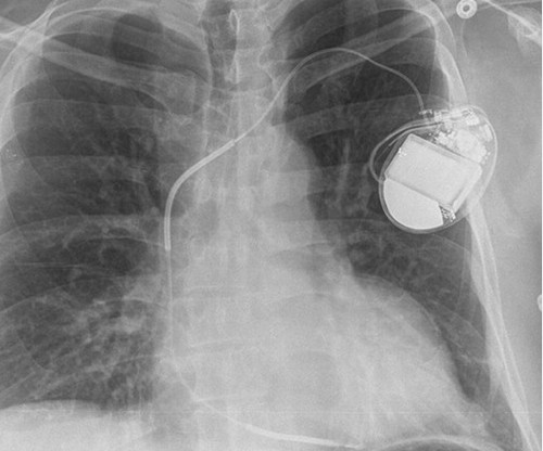
No evidence for ischemia jumps out. This is the shock coil and identifies this device as a defibrillator. CRT-D is cardiac resynchronization therapy with defibrillation capability, like the CXR above. CRT-P is cardiac resynchronization therapy with pacing only, without the ability to defibrillate. ECG 1 What do you think?
The ECG shows sinus rhythm with normal QRS complex morphology and significant subendocardial ischemia (SEI) pattern (ST depression in many leads, worst in lateral areas including leads II, V5-6, with reciprocal STE in aVR). Here is her ECG within 30 minutes of PCI: Improved, but still with ischemia. Pre-intervention. Post-intervention.
The possibility of an ischemic cause of the ventricular arrhythmia has to be considered! That said there were no clinical symptoms or ECG findings suggestive of ongoing ischemia. A workup was undertaken in search of a cause of the patient's ventricular arrhythmia. The idiopathic VTs are an interesting group of arrhythmias!
We organize all of the trending information in your field so you don't have to. Join thousands of users and stay up to date on the latest articles your peers are reading.
You know about us, now we want to get to know you!
Let's personalize your content
Let's get even more personalized
We recognize your account from another site in our network, please click 'Send Email' below to continue with verifying your account and setting a password.


Let's personalize your content