This site uses cookies to improve your experience. To help us insure we adhere to various privacy regulations, please select your country/region of residence. If you do not select a country, we will assume you are from the United States. Select your Cookie Settings or view our Privacy Policy and Terms of Use.
Cookie Settings
Cookies and similar technologies are used on this website for proper function of the website, for tracking performance analytics and for marketing purposes. We and some of our third-party providers may use cookie data for various purposes. Please review the cookie settings below and choose your preference.
Used for the proper function of the website
Used for monitoring website traffic and interactions
Cookie Settings
Cookies and similar technologies are used on this website for proper function of the website, for tracking performance analytics and for marketing purposes. We and some of our third-party providers may use cookie data for various purposes. Please review the cookie settings below and choose your preference.
Strictly Necessary: Used for the proper function of the website
Performance/Analytics: Used for monitoring website traffic and interactions
Coronary artery spasm (CAS), or Prinzmetal angina, is a recognised cause of myocardial ischaemia in non-obstructed coronary arteries which typically presents with anginal chestpain. The patient presented with recurrent palpitations and pre-syncope, with no chestpain.
Sent by anonymous, written by Pendell Meyers, reviewed by Smith and Grauer A man in his 40s presented to the ED with HTN, DM, and smoking history for evaluation of acute chestpain. He was eating lunch when he had sudden onset chest pressure, 9/10, radiating to his back, with sweating and numbness in both hands.
No ChestPain, but somnolent. The fact that this is syncope makes give it a far lower pretest probability than chestpain, but it was really more than syncope, as the patient actually underwent CPR and had hypotension on arrival of EMS. Here is the ED ECG (a photo of the paper printout) What do you think?
Written by Willy Frick A man in his 50s with history of hypertension, hyperlipidemia, and a 30 pack-year smoking history presented to the ER with 1 hour of acute onset, severe chestpain and diaphoresis. His ECG is shown: What do you think? What do you think? Given the R-R interval = 1160 msecs.
The primary endpoint was the incidence of device syndrome, a composite of patient-reported symptoms (chestpain, palpitations, migraines, dyspnea, and rash).Results:Of No significant differences were observed in documented arrhythmias, bleeding, or stroke. Nickel hypersensitivity was assessed using skin patch testing.
I went to the patient's chart: Elderly woman with stuttering chestpain and SOB, and dizziness. Tall R wave in lead V1 and/or early transition in the chest leads ( reflecting increased "septal" forces ). WPW Cardiac arrhythmias ( including AFib ). What do you think now? Abnormal ST-T wave abnormalities.
Whenever a patient does not have chestpain, the pre-test probability of OMI is diminished. Of course SOB, jaw pain, shoulder pain, etc can be a result of OMI, but the pretest probability is less and so you must scrutinize further. Lethal arrhythmias may be a terminal event ( VT/VFib; Asystole ).
Case sent by Logan Stark MD, written by Pendell Meyers A woman in her 70s presented with acute chestpain. Or perhaps the group beating is simply the result of sinus arrhythmia that by chance results in alternating R-R intervals of very similar size? No prior ECG was available. Here is her triage ECG: What do you think?
A middle-aged patient with lung cancer had presented to clinic complaining of generalized malaise, cough, and chestpain. Symptoms other than chestpain (malaise, cough in a cancer patient) 2. Inclusion criteria were chestpain, at least 2 serial cTnI in 24 hours, sinus rhythm , and at least 1 ECG.
A previously healthy middle-aged male presented shortly after the acute onset of chestpain very shortly before calling 911. On arrival, he was pain free: What do you think? Jerry Jones commented: "Any ST depression on the ECG of a patient with chestpain credible for ACS represents a reciprocal change until proved otherwise."
Written by Jesse McLaren A previously healthy 60 year old developed exertional chestpain with diaphoresis, and called EMS. There’s sinus arrhythmia with normal conduction, normal axis and normal voltages. Here’s the EMS ECG, digitized with PM cardio. What do you think? There’s loss of R waves in V2-3 with hyperacute waves V1-5.
Shortly after receiving epinephrine, the patient developed new leg cramps and chestpain. The chestpain was described as sharp and radiated to both arms. During active chestpain an ECG was recorded: Meyers ECG interpretation: Sinus tachycardia, normal QRS complex, STD in V2-V6, I, II, III and aVF.
A 50-something presented with acute chestpain. MY Thoughts on today’s “quick-look ECG” are the following: There is sinus arrhythmia. Here is her ED ECG. It was texted to me while I was out and about. He wrote: "Steve, what do you think about hyperacute T waves in this? 54-year-old female with CP. What do you think, Dear Reader?
The patient presented due to chestpain that was typical in nature, retrosternal and radiating to the left arm and neck. He denied any exertional chestpain. It is unclear if the patient was pain free at this time. He has a medical hx notable for hypertension, hyperlipidemia and previous tobacco use disorder.
The computer called it a normal ECG Algorithm unknown Aside : [There is some "sinus arrhythmia", which is indeed a normal finding. Sinus arrhythmia is sinus rhythm whose rate varies with respiration. If the longest P-P interval is 120 ms greater than the shortest, it is sinus arrhythmia. Burning pain subxiphoid and into throat."
Currently, he has no complaints: no palpitations, no shortness of breath, no syncope, no chestpain. There is a non-respiratory sinus arrhythmia present, which is essentially the minimal variant of a sick sinus syndrome. This is the ECG of an 81-year-old man with hypertension.
Case written and submitted by Ryan Barnicle MD, with edits by Pendell Meyers While vacationing on one of the islands off the northeast coast, a healthy 70ish year old male presented to the island health center for an evaluation of chestpain. The chestpain started about one hour prior to arrival while bike riding.
Written by Jesse McLaren, with edits from Smith and Grauer A 60 year old with no past medical history presented with two hours of chestpain radiating to the left arm, with normal vitals. Unfortunately, the reality is — that many ( most ) WPW patients who present with chestpain do not manifest intermittent preexcitation.
There is a patient with persistent chestpain and an initial troponin I over 52 ng/L; 52 ng/L has an approximate 70% PPV for acute type I MI in a chestpain patient. Pain was severe and persistent. CT angiography chest assessing for PE and dissection negative. Heparin drip was initiated. Is there STEMI?
It is from a 50-something with chestpain: What do you think? In my opinion — AI is not yet "there" with regard to interpretation of complex cardiac arrhythmias. I like to start my ECG assessment in patients with new chestpain by looking for at least 1 or 2 leads that I know are definitely abnormal.
The patient has acute chestpain. Instead — my thoughts were as follows: The rhythm is sinus , with marked bradycardia and a component of sinus arrhythmia. Tall R wave in lead V1 and/or early transition in the chest leads ( reflecting increased "septal" forces ). WPW Cardiac arrhythmias ( especially AFib ).
Written by Pendell Meyers A woman in her 50s presented with acute chestpain and lightheadedness since the past several hours. For our readers who enjoy the challenge of interpreting cardiac arrhythmias — today’s case offers a “gold mine” of PEARLS regarding the recognition of AV Wenckebach.
The word arrhythmia comes from two Greek words. So arrhythmia literally means absence or loss of rhythm. A cardiac arrhythmia therefore means loss of cardiac rhythm. These include breathlessness, chestpain, dizziness or even blackouts. appeared first on Dr Sanjay Gupta Cardiologist.
In the evening, a middle-aged man complained of chestpain at the nursing home. His chestpain was vague. He mentioned "cancer" and "chest". I’ll focus my comments on arrhythmia diagnosis. P EARL # 2 : In my experience — the most commonly overlooked arrhythmia ( by far! ) Fluids were started.
My THOUGHTS on ECG #1: We are told that the patient in today’s case had an episode of severe chestpain 3 nights prior to admission. This should include details regarding the dosing regimen — the nature of the ventricular arrhythmia being treated — and the patient’s “baseline” QTc at steady state for his/her current Sotalol dose.
Currently, he has no complaints: no palpitations, no shortness of breath, no syncope, no chestpain. There is a non-respiratory sinus arrhythmia present, which is essentially the minimal variant of a sick sinus syndrome. This is the ECG of an 81-year-old man with hypertension.
She was hemodynamically stable — and did not have chestpain, lightheadedness or syncope. With P waves labeled — Isn't it now much easier to appreciate that the atrial rhythm is quite regular ( with no more than a slight sinus arrhythmia )? For those with a special interest in cardiac arrhythmias — READ ON! —
This was written by Magnus Nossen, from Norway, with comments and additions by Smith A 50 something smoker with no previous medical hx contacted EMS due to acute onset chestpain. Upon EMS arrival the patient appeared acutely ill and complained of chestpain. Of academic interest — are the arrhythmias that developed.
BackgroundAmiodarone is a class III antiarrhythmic drug that is commonly used in the clinic to treat ventricular arrhythmias and atrial fibrillation. Despite normal annual check-ups, she developed abnormal liver and thyroid function tests, and imaging revealed lung and liver changes suggestive of amiodarone toxicity.
Note the Timed Contents that I detail below facilitate finding specific material. == ECG Podcast #4 — All About Comparison ECGs for 12-Leads and Arrhythmias ( Comparing ECGs seems so "easy" to do — but so often is not done correctly! ) — published by Mayo Clinic CV Podcast Series on 5/21/2024 ( 35 minutes ). What are the problems?
Patients may feel a fluttering in the chest, chestpain, shortness of breath and dizziness or lightheadedness as a result. It represents a unique opportunity to advance our therapy to the next level and potentially be able to treat patients with ventricular arrhythmias in a better way, without destroying healthy heart tissue.
And regarding arrhythmias: For more on the regular WCT — See My Comment in the May 5, 2020 post and in the April 15, 2020 post in Dr. Smith's ECG Blog. 11:35 — My views on: Will the computer ever be able to interpret complex arrhythmias? 14:45 — Using my definition — Are YOU an “ expert ” ECG interpreter? What are the problems?
We have long known that pulsed field ablation could open up an entirely new frontier in how we treat people battling the most complex cardiac arrhythmias. In addition to upcoming procedures in markets across Asia Pacific and Europe, Abbott anticipates approval for its U.S. chief medical officer of Abbott's electrophysiology business.
A 90 yo with a history of orthostatic hypotension had a near syncopal event followed by chestpain. Chestpain was resolved upon arrival in the ED. Idioventricular rhythm is a common "reperfusion arrhythmia." Exactly how they relate to ischemia, chestpain, and reperfusion can only be speculated about.
Just as important is pretest probability: did the patient report chestpain prior to collapse? After cardiac arrest, I ALWAYS wait 15 minutes after an ECG like this and record another. The ST depression usually resolves, or is clearly resolving (getting much better). Then assume there is ACS.
Circulation: Arrhythmia and Electrophysiology, Ahead of Print. Colchicine did not prevent atrial arrhythmia recurrence at 2 weeks (31% versus 32%; hazard ratio [HR], 0.98 [95% CI, 0.59–1.61];P=0.92) Postablation chestpain consistent with pericarditis was reduced with colchicine (4% versus 15%; HR, 0.26 [95% CI, 0.09–0.77];P=0.02)
Electrocardiogram in clinic showed sinus arrhythmia with early repolarization and no ischemic changes. Treadmill exercise stress test showed excellent functional capacity without exercise-induced chestpain or ischemic ECG changes. Invasive coronary angiography ruled out luminal narrowing or dynamic compression.
No chestpain. In a previously healthy adolescent ( who is 15 years old in today's case ) — the presentation of an acute febrile illness that is without a complaint of chestpain, is highly unlikely to be due to an acute MI. He was hemodynamically stable. How would YOU interpret the ECG in Figure-1 ?
He arrived to the ED by helicopter at 1507, about three hours after the start of his chestpain while chopping wood around noon. He arrived to the ED by ambulance at 1529, only a half hour after the start of his chestpain around 1500 while eating. Patient 2 was seen immediately after patient 1 by the same cardiologist.
Arrhythmias (Abnormal Heart Rhythms) Stress hormones can disrupt the signals that regulate your heartbeat, leading to arrhythmias – abnormal heart rhythms that cause your heart to beat too fast, too slow, or irregularly.
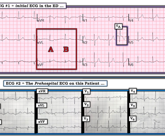
This is the prehospital ECG from an 81 year old man with acute chestpain. Arrhythmia? Today’s case recalled that scenario for me, in that it features recognition of an arrhythmia that fooled ED staff into thinking the ECG was showing an acute infarction. The medics did NOT activate the cath lab. Would you give lytics?
The ECG in Figure-1 was obtained from a previously healthy older man — who complained of chestpain and “lightheadedness” while this tracing was recorded. His chestpain had begun the night before. Figure-1: 12-lead ECG and long lead II rhythm strip — obtained from an older man with chestpain and “lightheadedness. (
Later, I found old ECGs: 5 month prior in clinic: V5 and V6 look like OMI 9 months prior in clinic with no chest symptoms: V5 and V6 look like OMI 1 year prior in the ED with chestpain: V5 and V6 sure look like a STEMI For this ECG and chestpain in the ED, the Cath lab activated. But the angiogram was clean.
The presenting complaint was chestpain — and the patient collapsed soon after arrival in the ED. C ASE F ollow- U p : As noted in today’s case presentation — this patient presented with chestpain, and then collapsed soon after the ECG in Figure-1 was obtained. Do YOU agree with this cardiologist ?
We organize all of the trending information in your field so you don't have to. Join thousands of users and stay up to date on the latest articles your peers are reading.
You know about us, now we want to get to know you!
Let's personalize your content
Let's get even more personalized
We recognize your account from another site in our network, please click 'Send Email' below to continue with verifying your account and setting a password.


Let's personalize your content