This site uses cookies to improve your experience. To help us insure we adhere to various privacy regulations, please select your country/region of residence. If you do not select a country, we will assume you are from the United States. Select your Cookie Settings or view our Privacy Policy and Terms of Use.
Cookie Settings
Cookies and similar technologies are used on this website for proper function of the website, for tracking performance analytics and for marketing purposes. We and some of our third-party providers may use cookie data for various purposes. Please review the cookie settings below and choose your preference.
Used for the proper function of the website
Used for monitoring website traffic and interactions
Cookie Settings
Cookies and similar technologies are used on this website for proper function of the website, for tracking performance analytics and for marketing purposes. We and some of our third-party providers may use cookie data for various purposes. Please review the cookie settings below and choose your preference.
Strictly Necessary: Used for the proper function of the website
Performance/Analytics: Used for monitoring website traffic and interactions
However, he suddenly developed a series of malignant ventricular arrhythmias. Below are printouts of some of the arrhythmias recorded. This time, the arrhythmia did not spontaneously terminate — but rather degenerated to VFib, requiring defibrillation. The arrhythmia starts with a PVC having a short coupling interval.
Abnormal readings can signal issues with circulation or lung function, prompting further investigation. Early detection of conditions like AFib, bradycardia, or tachycardia allows patients to address issues before they become critical. Sleep Monitoring Quality sleep is essential for heart health.
There are three mechanisms of arrhythmia: automatic, re-entry, and triggered. The most common triggered arrhythmia is Torsades de Pointes. It is a benign arrhythmia which requires no specific treatment. Possible mechanisms of ventricular arrhythmias elicited by ischemia followed by reperfusion. Do not treat AIVR.
Circulation: Cardiovascular Imaging. Other Arrhythmias ( PACs, PVCs, AFib, Bradycardia and AV conduction disorders — potentially lethal VT/VFib ). NOTE: Prediction of cardiac contusion "severity" on the basis of cardiac arrhythmias and ECG findings — is an imperfect science. 2015, March 1). Cramer, M. 2003, May).
Looking first at the long-lead II rhythm strip — there is significant bradycardia , with a heart R ate just under 40/minute. But the point to emphasize — is that it should only take seconds to recognize that there is bradycardia from significant AV block. = Would you approve her for a nonemergent surgical procedure?
Whatever the specific etiology of today's arrhythmia is, the “good news” is — that this rhythm will most probably improve with reperfusion of the "culprit" artery. That said — I found today's arrhythmia fascinating, and worthy of more in-depth analysis. Using calipers facilitates the process.
Circulation: Arrhythmia and Electrophysiology, Ahead of Print. Serious AEs were rare; 1 patient in the etripamil arm experienced transient severe bradycardia and syncope, assessed as due to hyper-vagotonia.Conclusions:Intranasal etripamil 70 mg reduced VR and improved symptom-relief and treatment-satisfaction.
VT is the second most common presenting arrhythmia. Vaso or inotropic medications are not harmless, and can precipitate life threatening arrhythmias. The physiologic reason for this — is thought to be the result of momentarily increased circulation from mechanical contraction arising from the "sandwiched in" QRS complex.
Dr. Smith circulated the initial ECG in today's case ( that I reproduce in Figure-1 ) to our group — initially without him yet knowing anything about this patient. I added, "Makes me wonder if this could be myocarditis in a younger adult — maybe even with sinus arrhythmia." Figure-1: The initial ECG in today's case.
Baseline bradycardia in endurance athletes limits the use of ß-blockers. The Role of Sinus Arrhythmia: I found it interesting to compare the long lead II rhythm strips in the 3 serial tracings from today’s case ( Figure-1 ). Note fairly marked irregularity of the R-R interval — indicative of definite sinus arrhythmia.
There is also bradycardia. Bradycardia puts patients at risk for "pause-dependent" Torsades de Pointes. Torsades in acquired long QT is much more likely in bradycardia because the QT interval following a long pause is longer still. malignant ventricular arrhythmias are present), rapid replacement of potassium is required.
Athlete’s bradycardia due to increased parasympathetic tone and decreased sympathetic tone is a well-known observation. Though sinus bradycardia is usual, other abnormalities like sinus arrhythmia, sinus arrest, wandering atrial pacemaker and coronary sinus rhythm have been described. Circulation. Heart Lung Circ.
Prior to Mizusawa's study, it was thought that the incidence of syncope, arrhythmia, or SCD in this cohort was low [7]. In light of the risk of arrhythmia events observed in the Mizusawa trial, a formal EP study might be reasonable to obtain in those with fever induced asymptomatic Brugada ECG changes to help risk stratify these patients.
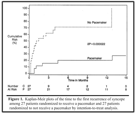
PVCs N ot generally considered abnormal ECG findings: Isolated PAC, First Degree AV Block, Sinus bradycardia at a rate of 35-45, and Nonspecific ST-T abnormalities (even if different from a previous ECG). Thus, if there is documented sinus bradycardia, and no suspicion of high grade AV block, at the time of the syncope, this is very useful.
A repeat ECG was performed as adult cardiology was asked to evaluate the patient for emerget PCI: Sinus bradycardia with persistent elevation in the inferior leads with reciprocal depression in aVL Patient was taken to cath lab with adult cardiology which revealed normal coronary arteries without evidence of occlusion MI. Circulation.
The possibility of an ischemic cause of the ventricular arrhythmia has to be considered! Below in Figure-5 is a 10-minute continuous lead II recording on oral Flecainide, now showing sinus bradycardia without a single PVC! A workup was undertaken in search of a cause of the patient's ventricular arrhythmia. No PVCs are seen.
Perhaps because the bradycardia in vasovagal syncope is only one part of the autonomic response. Phase 4 block is also referred to as "bradycardia dependent block." Circulation , 143 (10), 10621065. A stunning result. One of many examples in medical history that remind us of the importance of blinding in clinical trials.
We organize all of the trending information in your field so you don't have to. Join thousands of users and stay up to date on the latest articles your peers are reading.
You know about us, now we want to get to know you!
Let's personalize your content
Let's get even more personalized
We recognize your account from another site in our network, please click 'Send Email' below to continue with verifying your account and setting a password.


Let's personalize your content