This site uses cookies to improve your experience. To help us insure we adhere to various privacy regulations, please select your country/region of residence. If you do not select a country, we will assume you are from the United States. Select your Cookie Settings or view our Privacy Policy and Terms of Use.
Cookie Settings
Cookies and similar technologies are used on this website for proper function of the website, for tracking performance analytics and for marketing purposes. We and some of our third-party providers may use cookie data for various purposes. Please review the cookie settings below and choose your preference.
Used for the proper function of the website
Used for monitoring website traffic and interactions
Cookie Settings
Cookies and similar technologies are used on this website for proper function of the website, for tracking performance analytics and for marketing purposes. We and some of our third-party providers may use cookie data for various purposes. Please review the cookie settings below and choose your preference.
Strictly Necessary: Used for the proper function of the website
Performance/Analytics: Used for monitoring website traffic and interactions
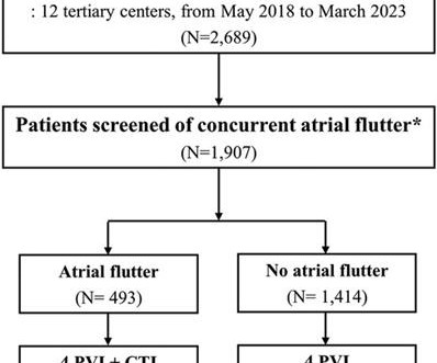
Typical atrialflutter commonly occurs in patients with atrial fibrillation (AF). Limited information exists regarding the effects of concurrent atrialflutter on the long-term outcomes of rhythm control. Patients who were screened for typical atrialflutter were included in the analysis ( n = 1,907).
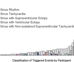
Sinus tachycardia – sinus rhythm above 100 bpm is a sinus tachycardia. Ventricular tachycardia – more than 7 consecutive complexes originating from ventricles at a rate of > 100 bpm. Supraventricular tachycardia – more than 7 consecutive complexes of supraventricular beats at a rate of > 100 bpm.
She had a single chamber ICD/Pacemaker implanted several years prior due to ventricular tachycardia. The ECG was interpreted as showing atrialflutter with 2:1 conduction. Answer : The ECG above shows a regular wide complex tachycardia. The heart rate could be compatible with that of a 2:1 conducted atrialflutter.
We reported the case of a 51-year-old woman who experienced multiple types of arrhythmias over three decades and was diagnosed with Danon disease late by genetic testing. Case summary A 51-year-old woman with a 36-year history of intermittent palpitations was admitted due to hemodynamically stable ventricular tachycardia (VT).
Wild-type transthyretin amyloid cardiomyopathy (ATTRwt-CM) is often accompanied by atrial fibrillation (AF), atrialflutter (AFL), and atrialtachycardia (AT), which are difficult to control because beta-blockers and antiarrhythmic drugs can worsen heart failure (HF).
Background Cardiac arrhythmias have been observed among patients hospitalised with acute COVID-19 infection, and palpitations remain a common symptom among the much larger outpatient population of COVID-19 survivors in the convalescent stage of the disease. Participants were instructed to trigger the monitor for palpitations.
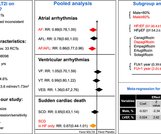
Objective We aimed to assess the effect of SGLT2i on arrhythmias by conducting a meta-analysis using data from randomized controlled trials(RCTs). Background Sodium-glucose co-transporter 2 inhibitors (SGLT2i) have shown cardioprotective effects via multiple mechanisms that may also contribute to decrease arrhythmias risk.
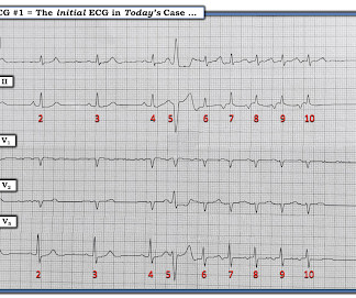
There is a regular wide complex tachycardia. A fully upright P-wave is typical atrial activity of atrialflutter as seen in V1. See these example cases of upright P-waves: Case Continued Thus, I was all but certain that this was atrialflutter. If it is flutter, it will reveal the underlying flutter waves.
The rhythm differential for narrow, regular, and tachycardic is sinus rhythm, SVT (encompassing AVNRT, AVRT, atrial tach, etc), and atrialflutter (another supraventricular rhythm which is usually considered separately from SVTs). Therefore this patient is either in some form of SVT or atrialflutter.
Unlike paroxysmal AF, which describes symptoms that last for seven days or fewer, persistent AF is a sustained arrhythmia that lasts for more than a week 1. Early treatment of persistent AF can reduce the risk of blood clots, stroke, and heart failure, and may prevent the disease from becoming permanent. The company now anticipates U.S.
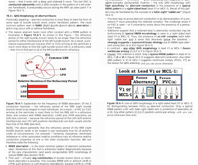
Wide-complex tachycardia: VT or aberrant, or "other?" The patient had a history of paroxysmal atrial fibrillation and several cardioversions. A wide-complex tachycardia in an older patient must immediately suggest ventricular tachycardia. Instead, the rate of 150, plus the history of AF, suggested atrialflutter.
A series of cardiac arrhythmias were seen during the course of her resuscitation — including the interesting arrhythmia shown in the long lead II of Figure-1. PEARL # 3: At this point — the most time-efficient step for solving today's rhythm will be to determine the nature of atrial activity.
What arrhythmia is present? Let's first consider the heart rate: with a heart rate of 194 beats/min, the heart rate is too low for atrialflutter (1:1) (except in patients who have been pre-treated with medication), and the rate would be unusually high for atrialflutter with 2:1 conduction.
Initial ECG in the ED: Presenting ECG : Wide-complex tachycardia at a rate about 200. This is overwhelmingly likely to be ventricular tachycardia, even if only age and medical history are considered. Nevertheless, the widths of both the QRS complex and the RS duration are similar in both the old ECG and the tachycardia.
Cheng and Zhang to our paper1, we note that despite ARVC being a relatively uncommon cardiomyopathy, we uniquely report on a large clinical experience with very long-term follow-up after VT ablation and confirm a high incidence of atrialflutter (AFL) and the effectiveness and low risk of catheter ablation of AFL.
This strip was obtained: Apparent Wide Complex Tachycardia at a rate of 280 What do you think? To me, it was clearly atrialflutter with 1:1 conduction. The rate of 280 is just right for atrialflutter. The waves look like atrialflutter waves, NOT like a wide ventricular complex.
She was noted to be in heart failure, with hypotension and tachycardia. With decompensated heart failure and adequate prior anticoagulation, the patient underwent direct current synchronised cardioversion for her arrhythmia. Echocardiography demonstrated severe calcific mitral stenosis with pulmonary hypertension.
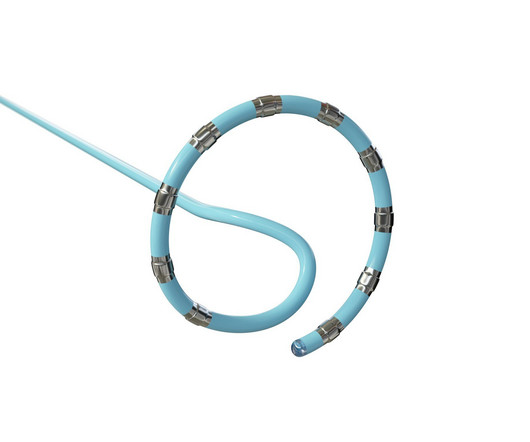
Although the QDOT MICRO™ Catheter was mainly designed for pulmonary vein isolation (PVI) its versatility to treat atrial fibrillation (AF) and other types of arrhythmias was recently evaluated by the FAST and FURIOUS study series and other studies and will be presented in this article.
There is a narrow complex tachycardia at a rate of 130. ECGs: there is a regular narrow complex tachycardia still at a rate of exactly 130, with no P-waves and also no change since the prehospital ECG. Leads II and aVF appear to have flutter waves. I diagnosed atrialflutter with 2:1 conduction. Is is sinus?
First, we have a narrow-complex, regular tachycardia, with a rate of about 135-140. This narrows our differential for the rhythm down to sinus tachycardia, paroxysmal supraventricular tachycardia (PSVT, or SVT), and atrialflutter. They are flutter waves, and the rhythm is 2:1 atrialflutter.
Since the most common ventricular response to untreated AFlutter is with 2:1 AV conduction — this results in a ventricular rate HALF as fast as the flutter rate in the atria — and 300 ÷ 2 ~150/minute ( usual range ~130-170/minute ). And a Final Tracing. Reviews PEARLS regarding the ECG diagnosis of AFlutter — and — What's "New"?
Here I put arrows: Arrows shows slow atrialflutter waves. Arrhythmia? Today’s case recalled that scenario for me, in that it features recognition of an arrhythmia that fooled ED staff into thinking the ECG was showing an acute infarction. These mimic ST Elevation. But there is no STE. Would you give lytics? You decide.
Introduction:Supraventricular tachycardia (SVT) is common and poorly tolerated in patients who have undergone Fontan procedure. AtrialTachycardia (70%) and Typical AtrialFlutter (65%) were the most common SVTs ablated. Circulation, Volume 150, Issue Suppl_1 , Page A4142266-A4142266, November 12, 2024.
In all probability, this dilation is a form of atrialtachycardia and atrial cardiomyopathy. However, underlying lesions such as hypertension, mitral valve disease, COPD, ASD, and TR greatly influence the degree of atrial enlargement. We know atrialflutters can be confined to one atrium.
The rhythm is indeed irregularly irregular, so atrial fibrillation must be considered. There are 5 other rhythms that are irregularly irregular , though atrial fibrillation is by far the most common: 1. Multifocal AtrialTachycardia 2. How can you avoid overlooking this arrhythmia? Sinus with multifocal PACs 3.
Abstract Introduction Atrial fibrillation and atrialflutter originating from the donor s heart is a commonly reported complication post heart transplant. Case A 47-year-old male presented with atrialtachycardia 6 months post heart transplant.
Circulation: Arrhythmia and Electrophysiology, Ahead of Print. BACKGROUND:Inflammation may promote atrial fibrillation (AF) recurrence after catheter ablation. Colchicine did not prevent atrialarrhythmia recurrence at 2 weeks (31% versus 32%; hazard ratio [HR], 0.98 [95% CI, 0.59–1.61];P=0.92) 2.02];P=0.89).
Electrical cardioversion is an atrial fibrillation medical procedure often recommended for patients experiencing irregular and rapid heartbeats. Cardioversion is used to correct abnormal heart rhythms, also known as arrhythmias. Atrialflutter: This is a rapid but regular heart rhythm often progressing to AFib.
What is unusual about this arrhythmia? Doing so suggests that the R-R interval of this exceedingly rapid arrhythmia is just a tiny amount over 1 large box — which corresponds to a ventricular rate just under 300/minute ( ie, between 290-300/minute ). PEARL # 3: AtrialFlutter with 1:1 AV conduction is rare!
We see a regular tachycardia with a narrow QRS complex and no evidence of OMI or subendocardial ischemia. The differential of a regular narrow QRS tachycardia is sinus tachycardia, SVT, and atrialflutter with regular conduction. There are no P waves preceding the QRS complexes, and no clear flutter waves.
By this definition, a variety of rhythms may qualify as “SVTs” — including sinus tachycardia, atrialflutter or fibrillation, MAT, AVRT/AVNRT, among others. Why Isn’t this a Run of AtrialTachycardia? — ECG Blog #185 — Reviews the P s, Q s, 3 R Approach to Arrhythmia Interpretation.
The principal d ifferential d iagnosis i s similar to what we derived in the October 16, 2019 Case : i ) Sinus Tachycardia ; ii ) Reentry SVT ( either A VNRT if the reentry circuit is contained within the normal AV nodal pathway — or A VRT if an accessory pathway is involved ) ; iii ) AtrialTachycardia ; or iv ) AtrialFlutter.
Here is his 12-lead: There is a wide complex tachycardia with a rate of 257, with RBBB and LPFB (right axis deviation) morphology. Read about Fascicular VT here: Idiopathic Ventricular Tachycardias for the EM Physician Case Continued He was completely stable, so adenosine was administered. See Learning point 1 below. Arch Intern Med.
Atrial fibrillation, atrialtachycardia or atrialflutter with Wenckebach conduction. The meaning of this English proverb, " Birds of a Feather. " — is that people of similar type, interest or character tend to mutually associate.
Jimenez and Ali co-founded AccurKardia in 2019 with a vision for unlocking the value of the ECG signal, and the company currently markets one of the few FDA-cleared solutions for automated ECG interpretation and arrhythmia detection. Here’s the rest of their story and vision for ECG automation.
Here was his ED ECG: There is sinus tachycardia (rate about 114) with nonspecific ST-T abnormalities. There is a large peaked P-wave in lead II (right atrial enlargement) There is left axis deviation consistent with left anterior fascicular block. See my quick review of atrialtachycardia below) The tachycardia spontaneously resolved.
a global leader in cardiac arrhythmia treatment and part of Johnson & Johnson MedTech , today announced European CE mark approval of the VARIPULSE Platform for the treatment of symptomatic drug refractory recurrent paroxysmal atrial fibrillation ( AF ) using pulsed field ablation (PFA). [ii]
If the patient has Abnormal Vital Signs (fever, hypotension, tachycardia, or tachypnea, or hypoxemia), then these are the primary issue to address, as there is ongoing pathology which must be identified. The most recent and probably best study is this: Canadian Syncope Arrhythmia Risk Score. Vasovagal syncope is generally benign.
Additionally, the patient had no other apparent reason to have sinus tachycardia (such as volume depletion, bleeding, fever). So the most likely rhythm in ECG 1 is ectopic atrialtachycardia. Therefore the first part of ECG 1 shows ectopic atrialtachycardia with biventricular pacing. Point 1: What is PVARP?
During observation in the ED the patient had multiple self-terminating runs of Non-Sustained monomorphic Ventricular Tachycardia (NSVT). The possibility of an ischemic cause of the ventricular arrhythmia has to be considered! A workup was undertaken in search of a cause of the patient's ventricular arrhythmia. No PVCs are seen.
Methods and Results ATA recurrence was defined as 30s recurrence of atrial fibrillation, atrialflutter or atrialtachycardia after a 90-day blanking period and through 12-months. Univariate and multivariable Cox regression analysis (with ATA recurrence as an endpoint) was performed to identify CBA responders.
We organize all of the trending information in your field so you don't have to. Join thousands of users and stay up to date on the latest articles your peers are reading.
You know about us, now we want to get to know you!
Let's personalize your content
Let's get even more personalized
We recognize your account from another site in our network, please click 'Send Email' below to continue with verifying your account and setting a password.


Let's personalize your content