This site uses cookies to improve your experience. To help us insure we adhere to various privacy regulations, please select your country/region of residence. If you do not select a country, we will assume you are from the United States. Select your Cookie Settings or view our Privacy Policy and Terms of Use.
Cookie Settings
Cookies and similar technologies are used on this website for proper function of the website, for tracking performance analytics and for marketing purposes. We and some of our third-party providers may use cookie data for various purposes. Please review the cookie settings below and choose your preference.
Used for the proper function of the website
Used for monitoring website traffic and interactions
Cookie Settings
Cookies and similar technologies are used on this website for proper function of the website, for tracking performance analytics and for marketing purposes. We and some of our third-party providers may use cookie data for various purposes. Please review the cookie settings below and choose your preference.
Strictly Necessary: Used for the proper function of the website
Performance/Analytics: Used for monitoring website traffic and interactions
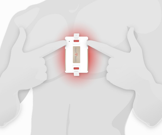
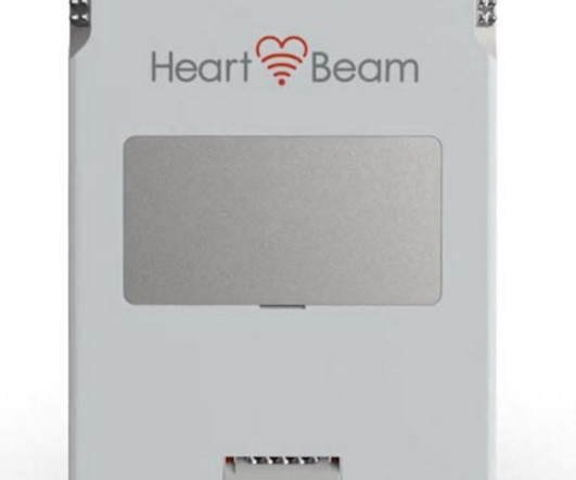
a medical technology company focused on transforming cardiac care through the power of personalized insights, today announced new study data demonstrating that HeartBeam AI combined with vectorcardiography (VCG) outperformed an expert panel of heart rhythm cardiologists in detecting atrialflutter. The data was presented by Joshua M.
The data was presented by Vivek Reddy , MD, Director of Cardiac Arrhythmia Services at The Mount Sinai Hospital , during the European Heart Rhythm Association (EHRA) conference in Berlin, Germany. for single-lead ECG) without sacrificing the ability to identify those individuals without atrialflutter (specificity of 98.7%
To me, it was clearly atrialflutter with 1:1 conduction. The rate of 280 is just right for atrialflutter. The waves look like atrialflutter waves, NOT like a wide ventricular complex. Reverted to atrial fibrillation with RVR while in the hospital 3 times and needed cardioversion.
Circulation: Arrhythmia and Electrophysiology, Ahead of Print. BACKGROUND:Inflammation may promote atrial fibrillation (AF) recurrence after catheter ablation. Colchicine did not prevent atrialarrhythmia recurrence at 2 weeks (31% versus 32%; hazard ratio [HR], 0.98 [95% CI, 0.59–1.61];P=0.92) 2.02];P=0.89).
Instead, the rate of 150, plus the history of AF, suggested atrialflutter. A close inspection of lead II showed P or flutter waves at a rate of about 300 bpm, also supporting atrialflutter. There appear to be flutter waves at a rate of 300. Flecainide encourages new atrialflutter.
Atrial fibrillation (AF) is the most common sustained arrhythmia and associated with increased morbidity and mortality. PubMed was queried for entries on AF and rurality: (atrial fibrillation OR atrialflutter) AND (rural OR urban OR rurality OR metro OR metropolitan) AND (united states OR US OR U.S.)
The ECG was interpreted as showing atrialflutter with 2:1 conduction. The heart rate could be compatible with that of a 2:1 conducted atrialflutter. Also, lead I could give the initial impression of showing flutter waves. On her 5th hospital day — she was given Amiodarone, which successfully converted the rhythm.
Electrical cardioversion is an atrial fibrillation medical procedure often recommended for patients experiencing irregular and rapid heartbeats. Cardioversion is used to correct abnormal heart rhythms, also known as arrhythmias. Atrialflutter: This is a rapid but regular heart rhythm often progressing to AFib.
The 12-lead ECG and long lead II rhythm strip shown in Figure-1 — was obtained from a previously healthy, elderly woman who collapsed in the hospital parking lot. A series of cardiac arrhythmias were seen during the course of her resuscitation — including the interesting arrhythmia shown in the long lead II of Figure-1.
The differential of a regular narrow QRS tachycardia is sinus tachycardia, SVT, and atrialflutter with regular conduction. There are no P waves preceding the QRS complexes, and no clear flutter waves. This includes sinus tachycardia, atrial fibrillation or flutter, MAT, and others.
During the 5-year follow up period, 13 (59%) patients with follow up had cardiovascular (CV) hospitalization and 1 patient died. Atrial Tachycardia (70%) and Typical AtrialFlutter (65%) were the most common SVTs ablated. Rate of recurrence did not differ between those who had the procedure before or after 2018.
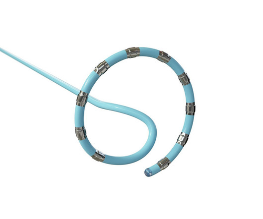
a global leader in cardiac arrhythmia treatment and part of Johnson & Johnson MedTech , today announced European CE mark approval of the VARIPULSE Platform for the treatment of symptomatic drug refractory recurrent paroxysmal atrial fibrillation ( AF ) using pulsed field ablation (PFA). [ii]
The patient was given furosemide and admitted to the hospital. There is atrial activity before every QRS, but that activity has negative polarity, so it is not sinus rhythm. The other atrialflutter types are: 1. A bedside POC cardiac ultrasound was done: Findings: Decreased left ventricular systolic function.
Finally, much of this correlates well with The new Canadian Syncope Arrhythmia Risk Score , just published in 2016, results of which are given below in the Annotated Bibliography. The most recent and probably best study is this: Canadian Syncope Arrhythmia Risk Score. Vasovagal syncope is generally benign. Thiruganasambandamoorthy, V.,
We organize all of the trending information in your field so you don't have to. Join thousands of users and stay up to date on the latest articles your peers are reading.
You know about us, now we want to get to know you!
Let's personalize your content
Let's get even more personalized
We recognize your account from another site in our network, please click 'Send Email' below to continue with verifying your account and setting a password.


Let's personalize your content