This site uses cookies to improve your experience. To help us insure we adhere to various privacy regulations, please select your country/region of residence. If you do not select a country, we will assume you are from the United States. Select your Cookie Settings or view our Privacy Policy and Terms of Use.
Cookie Settings
Cookies and similar technologies are used on this website for proper function of the website, for tracking performance analytics and for marketing purposes. We and some of our third-party providers may use cookie data for various purposes. Please review the cookie settings below and choose your preference.
Used for the proper function of the website
Used for monitoring website traffic and interactions
Cookie Settings
Cookies and similar technologies are used on this website for proper function of the website, for tracking performance analytics and for marketing purposes. We and some of our third-party providers may use cookie data for various purposes. Please review the cookie settings below and choose your preference.
Strictly Necessary: Used for the proper function of the website
Performance/Analytics: Used for monitoring website traffic and interactions
Food and Drug Administration (FDA) has granted 510(k) clearance for its first-of-a-kind, AI-powered AISAP CARDIO point-of-care ultrasound (POCUS) software platform. We know that structural heart disease and heart failure are the leading causes of hospitalization and morbidity in the U.S.
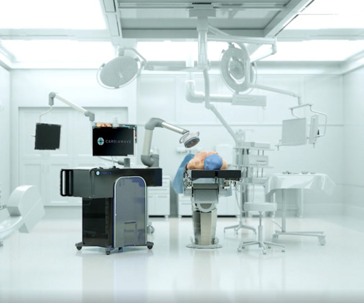
Presentation title: QUALITY OF LIFE ASSESSMENT AT 30-DAYS FOLLOW-UP OF THE VALVOSOFT PIVOTAL STUDY ON SEVERE AORTIC VALVE STENOSE PATIENTS" (control number 16930) Time and date: April 8, 2024 - 9:32 a.m.–9:42 A summary of the poster session will be published on the Journal of the American College of Cardiology’s website.
Fetal aortic valvuloplasty is considered for fetuses with severe valvar aorticstenosis and echocardiographic features suggesting a risk of progression to hypoplastic left heart syndrome. So if progression to hypoplastic left heart syndrome can be prevented by fetal aortic valvuloplasty, that would be theoretically a great boon.
510(k) clearance secured from FDA allows for EIQ’s AI-enabled solution, EchoSolv AS, to be marketed and sold in the USA Clearance marks a major milestone and will allow for rapid commercial scale-up EIQ is in advanced discussions with a range of US healthcare providers around the potential uptake of EchoSolv AS Working alongside US consultancy to obtain (..)
Few of them currently have the equipment and expertise to diagnose valvular heart disease, but recent studies have demonstrated that healthcare professionals can use a combination of portable ultrasound devices and AI to diagnose heart diseases as well.
24: Joint American College of Cardiology/Journal of the American College of Cardiology Late-Breaking Clinical Trials (Session 402) Saturday, April 6 9:30 – 10:30 a.m.
The Queen of Hearts disagrees, diagnosing OMI with high confidence: Case Continued: The EKG was not immediately recognized by the emergency provider, who ordered a CT scan to rule out aortic dissection at 1419. If it is angina, lowering the BP with IV Nitroglycerine may completely alleviate the pain and the (unseen) ECG ischemia.
Smith comment: This patient did not have a bedside ultrasound. Had one been done, it would have shown a feature that is apparent on this ultrasound (however, this patient's LV function would not be as good as in this clip): This is recorded with the LV on the right. Look at the aortic outflow tract. What do you see?
Although indexing effective orifice area (EOA) by body surface area (BSA) is recommended, this method has several disadvantages, since it corrects by acquired fatty tissue. Our aim was to analyze the value of.
What is the evidence for intra-aortic balloon pumps, percutaneous ventricular assist devices and ECMO in the patient with cardiogenic shock? What are the best strategies to efficiently get the patient in cardiogenic shock to definitive care, whether that be the cath lab or the operating room?
During medical school, one of the classic bedside exam questions we get is how to differentiate the valve issues of aorticstenosis and mitral regurgitation, which produce similar but different murmurs when you listen with a stethoscope. You use an ultrasound. When I teach trainee doctors, I ask them this very question.
They found non-obstructive CAD, with only a 20% stenosis of OM2 and 10% RCA. The next morning the patient went for his routine echocardiogram, where the operator noticed a dilated aortic root at 5.47 cm with severe aortic insufficiency. Beware a negative Bedside ultrasound. No acute culprit. He was admitted to cardiology.
History sounds concerning for ACS (could be critical stenosis, triple vessel), but differential also includes dissection, GI bleed, etc. 2 cases of AorticStenosis: Diffuse Subendocardial Ischemia on the ECG. His response: “subendocardial ischemia. Anything more on history? POCUS will be helpful.” Left main? 3-vessel disease?
Due to the severity of the pain and the high BP, they obtained an aortic dissection CT. Left main: no significant stenosis. LAD: proximal 60% eccentric stenosis the hemodynamic significance of which is indeterminate. RCA: Dominant: Mid 50-60% stenosis. At this point, the cath lab was activated.
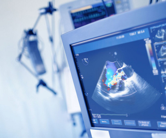
During echocardiography, a transducer transmits the ultrasound beam towards the heart. Opening and closing movements of the aortic and mitral valves are visible. Planimetry of mitral valve area can be obtained in parasternal short axis view in case of mitral stenosis. Exact position and angulation will vary between individuals.
An elderly patient with a ruptured abdominal aortic aneurysm: Formal ECG Interpretation (final read in the chart!) : "Inferior ST elevation, lead III, with reciprocal ST depression in aVL." A bedside ultrasound was done by the emergency physician, using Speckle Tracking. What do you think? Unfortunately, that video is unavailable.
Venn diagram highlighting the main similarities and differences between heart failure with preserved ejection fraction (HFpEF) and aorticstenosis with preserved ejection fraction (ASpEF). Patients with ASpEF eligible for transcatheter aortic valve replacement ( n = 125) also performed cardiac computed tomography (CT).
Aortic Dissection, Valvular (especially AorticStenosis), Tamponade. Check : [vitals, SOB, Chest Pain, Ultrasound] If the patient has Abdominal Pain, Chest Pain, Dyspnea or Hypoxemia, Headache, Hypotension , then these should be considered the primary chief complaint (not syncope). heart auscultation (aorticstenosis); c.
28, 2024 — Cardiawave SA, developer of Valvosoft Non-Invasive Ultrasound Therapy (NIUT) device for treating severe symptomatic calcific aorticstenosis (CAS), announced that the device met the primary endpoint in its pivotal study and improved or stabilized heart failure symptoms for 80.5 tim.hodson Thu, 11/07/2024 - 10:19 Oct.
I suspect pulmonary edema, but we are not given information on presence of B-lines on bedside ultrasound, or CXR findings. Smith : "decompensation" of aorticstenosis might have initiated this entire cascade. What "initiates" the aorticstenosis cascade? We certainly know that there is hypoxia.
We organize all of the trending information in your field so you don't have to. Join thousands of users and stay up to date on the latest articles your peers are reading.
You know about us, now we want to get to know you!
Let's personalize your content
Let's get even more personalized
We recognize your account from another site in our network, please click 'Send Email' below to continue with verifying your account and setting a password.


Let's personalize your content