This site uses cookies to improve your experience. To help us insure we adhere to various privacy regulations, please select your country/region of residence. If you do not select a country, we will assume you are from the United States. Select your Cookie Settings or view our Privacy Policy and Terms of Use.
Cookie Settings
Cookies and similar technologies are used on this website for proper function of the website, for tracking performance analytics and for marketing purposes. We and some of our third-party providers may use cookie data for various purposes. Please review the cookie settings below and choose your preference.
Used for the proper function of the website
Used for monitoring website traffic and interactions
Cookie Settings
Cookies and similar technologies are used on this website for proper function of the website, for tracking performance analytics and for marketing purposes. We and some of our third-party providers may use cookie data for various purposes. Please review the cookie settings below and choose your preference.
Strictly Necessary: Used for the proper function of the website
Performance/Analytics: Used for monitoring website traffic and interactions
Transcipt of video: Mild tricuspid regurgitation is often noted on echocadiogram reports and sometimes causes a little bit of worry and a lot of questions are asked on mild tricuspid regurgitation. What is this mild tricuspid regurgitation? And mild tricuspid regurgitation is just a small leak from the tricuspid valve.
Objective A novel artificial intelligence-based phenotyping approach to stratify patients with severe aortic stenosis (AS) prior to transcatheter aortic valve replacement (TAVR) has been proposed, based on echocardiographic and haemodynamic data. ±15.8 ±15.1 mm Hg, p value: 0.0079). to 84.7%) and 74.6% (95% CI 65.9%
Methods and Results This case report discusses a 65-year-old man who had previously undergone pulmonary vein isolation (PVI) and cavo-tricuspid isthmus ablation for atrial fibrillation before ASD closure, respectively. He developed atrial tachycardia (AT) and underwent catheter ablation.
The tricuspid valve is the right atrioventricular valve. The pulmonary semilunar valve is between the right ventricle and the pulmonary trunk. The aortic semilunar valve is between the left ventricle and the aorta. Like the heart chambers, there are four heart valves between each of the chambers.
The RFCs were much more successful at classifying murmurs from the pulmonary and tricuspid valves (AUROC = 0.83 and 0.78, respectively) when compared with the aortic and mitral valves (AUROC = 0.72 All RFC models were evaluated using the area under their receiver operating characteristic curves (AUROCs). and 0.65, respectively).
Usual structures imaged in this view are the right ventricular free wall and outflow region, interventricular septum, aorta, and aortic valve, left ventricular outflow tract, anterior and posterior mitral leaflets, left ventricular cavity, posterior wall of left ventricle and left atrium. Colour flow shows the flow in pulmonary artery.
We are blessed with 4 heart valves – 2 on the left side which are known as the mitral and aortic valves and 2 on the right side – the tricuspid and pulmonary valves.
Objectives This study aimed to evaluate the prognostic value of coronary microvascular dysfunction (CMD) at long term after transcatheter aortic valve implantation (TAVI) and to explore its relationship with extravalvular cardiac damage (EVCD). CMD was defined as IMR angio ≥30 units.
Assessment of fluid overload identifies aortic stenosis (AS) patients at high risk and treatment of fluid overload may potentially improve the post-interventional clinical course. TAVI, transcatheter aortic valve implantation. Right-sided cardiac damage (rCD) was defined as pulmonary vasculature/tricuspid/right ventricular damage.
Program Designations Access and Publications (A&P) 1 Participant User File (PUF) 2 Task Force on Funded Research (TFR) 3 Special Projects 4 Adult Cardiac Surgery Database Lead Author Title Publication Date William Keeling 2 National Trends in Emergency Coronary Artery Bypass Grafting European Journal of Cardiothoracic Surgery October 2023 Jake (..)
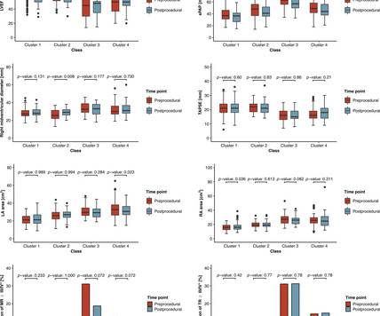
AF, atrial fibrillation; LAVI, left atrial volume index; RA, right atrial; RV, right ventricular; sPAP, systolic pulmonary artery pressure; SVI, stroke volume index; TR, tricuspid regurgitation. Aims Paradoxical low-flow, low-gradient aortic stenosis (pLFLG AS) may represent a diagnostic challenge, and its pathophysiology is complex.
We organize all of the trending information in your field so you don't have to. Join thousands of users and stay up to date on the latest articles your peers are reading.
You know about us, now we want to get to know you!
Let's personalize your content
Let's get even more personalized
We recognize your account from another site in our network, please click 'Send Email' below to continue with verifying your account and setting a password.


Let's personalize your content