This site uses cookies to improve your experience. To help us insure we adhere to various privacy regulations, please select your country/region of residence. If you do not select a country, we will assume you are from the United States. Select your Cookie Settings or view our Privacy Policy and Terms of Use.
Cookie Settings
Cookies and similar technologies are used on this website for proper function of the website, for tracking performance analytics and for marketing purposes. We and some of our third-party providers may use cookie data for various purposes. Please review the cookie settings below and choose your preference.
Used for the proper function of the website
Used for monitoring website traffic and interactions
Cookie Settings
Cookies and similar technologies are used on this website for proper function of the website, for tracking performance analytics and for marketing purposes. We and some of our third-party providers may use cookie data for various purposes. Please review the cookie settings below and choose your preference.
Strictly Necessary: Used for the proper function of the website
Performance/Analytics: Used for monitoring website traffic and interactions
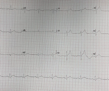
This ECG together with these symptoms is certainly concerning for OMI, but the ECG is not fully diagnostic, and another consideration could be acute pericarditis. Mistaking OMI for pericarditis is a much more harmful error than the converse. The rate is tachycardic, which is uncommon in OMI and common in pericarditis.
Overall, this looks like one of the rare ECGs that is actually specific for pericarditis in my opinion. Pericarditis maybe." Two weeks ago he had a significant MVC with many severe injuries, including aortic injury s/p endovascular repair. Meyers' words — "is one of the rare ECGs that is actually specific for pericarditis".
mm has been described in normal subjects) Overall impression: In my opinion and experience, this ECG most likely represents a normal baseline ECG, but with a small chance of pericarditis instead. I texted this to Dr. Smith without any information, and this was his reply: "This could be pericarditis but probably is normal variant."
Post op chest pain Typical of post-op pericarditis (postpericardiotomy syndrome) There is ST Elevation in II > III, and STE in V3-5, but with flat T-wave. The ST elevation is far more prominent than the T-wave and this is what I see as the defining feature differentiating OMI from pericarditis.
This is a value typical for a large subacute MI, n ormal value 48 hours after myocardial infarction is associated with Post-Infarction Regional Pericarditis ( PIRP ). As already mentioned, this patient could have post-infarction regional pericarditis from a large completed MI. Sinus tachycardia has many potential causes. Hammill SC.
When there is MI extending all the way to the epicardium (transmural), that infarcted epicardium is often inflamed (postinfarction regional pericarditis, or PIRP). What complication is the patient with post-infarction regional pericarditis at risk for? 3) Oliva et al. (4) Lessons : 1. One should be on the alert for myocardial rupture.
Another type of pain originating from the heart is due to an inflammation of its covering known as pericarditis. Pain of pericarditis can increase on swallowing and mimic pain from the food pipe. Pain of aortic dissection is tearing type and is most severe in the beginning. Pain may be relieved on leaning forwards.
A dissection flap is noted in the intrabdominal aorta, and the aortic outflow tract is also noted to appear wider than normal. The CT angio showed a type A aortic dissection extending from the aortic root proximally to the carotid and left subclavian artery and distally to the common femoral arteries.
ECG of pneumopericardium and probable myocardial contusion shows typical pericarditis Male in 30's, 2 days after Motor Vehicle Collsion, complains of Chest Pain and Dyspnea Head On Motor Vehicle Collision. : A Child with Blunt Trauma -- See how the ECG can be definite for myocardial contusion, but subtle, and what happens if you miss it.
Then the patient's pain then resolved spontaneously after 2 sublingual nitroglycerine and another ECG was recorded ECG 2 at 16 minutes ST ELEVATION CONSISTENT WITH INJURY, PERICARDITIS, OR EARLY REPOLARIZATION Overread same Smith : The T-waves are now MUCH smaller. They also wanted an aortic CT which was negative.
Pericarditis? There was concern for aortic dissection, so a CT was done and was negative. A straight ST segment virtually never happens in inferior ST elevation that is NOT due to OMI (normal variant, pericarditis) 4. 60-something with h/o MI and stents presented with chest pain radiating to the back and nausea/vomiting.
As always, takotsubo cardiomyopathy and focal pericarditis can mimic OMI, but takotsubo almost never mimics posterior MI, and both are diagnoses of exclusion after a negative cath. Chest pain and Concordant ST Depression in a patient with aortic valve and previously normal angiogram Right Bundle Branch Block and ST Depression in V1-V3.
ECG of pneumopericardium and probable myocardial contusion shows typical pericarditis Male in 30's, 2 days after Motor Vehicle Collsion, complains of Chest Pain and Dyspnea Head On Motor Vehicle Collision. ST depression. Myocardial Contusion?
In this ECG Cases blog we look at 10 cases of patients with chest pain, including false positive STEMI, false negative STEMI, and other causes to help hone your ECG interpretation skills in time-sensitive cases where those very ECG skills might save a life.
CT angiogram chest: no aortic dissection or pulmonary embolism. Pericarditis? He spent several days in the PICU, undergoing workup including: Serial troponins: rising from 5,700 ng/L (unknown if I or T) to greater than 25,000 ng/L (greater than the lab's upper limit of reporting). No further troponins were measured.
The exception is with postinfarction pericarditis , in which a completed transmural infarct results in inflammation of the subepicardial myocardium and STE in the distribution of the infarct, and which results in increased STE and large upright T-waves. These findings together are more commonly seen with pericarditis.
We organize all of the trending information in your field so you don't have to. Join thousands of users and stay up to date on the latest articles your peers are reading.
You know about us, now we want to get to know you!
Let's personalize your content
Let's get even more personalized
We recognize your account from another site in our network, please click 'Send Email' below to continue with verifying your account and setting a password.


Let's personalize your content