This site uses cookies to improve your experience. To help us insure we adhere to various privacy regulations, please select your country/region of residence. If you do not select a country, we will assume you are from the United States. Select your Cookie Settings or view our Privacy Policy and Terms of Use.
Cookie Settings
Cookies and similar technologies are used on this website for proper function of the website, for tracking performance analytics and for marketing purposes. We and some of our third-party providers may use cookie data for various purposes. Please review the cookie settings below and choose your preference.
Used for the proper function of the website
Used for monitoring website traffic and interactions
Cookie Settings
Cookies and similar technologies are used on this website for proper function of the website, for tracking performance analytics and for marketing purposes. We and some of our third-party providers may use cookie data for various purposes. Please review the cookie settings below and choose your preference.
Strictly Necessary: Used for the proper function of the website
Performance/Analytics: Used for monitoring website traffic and interactions
ObjectiveSpinal cord ischemia due to damage or occlusion of the orifices of aortic segmental arteries (ASA) is a serious complication of open and endovascular aortic repair. Furthermore, it aids in planning and conducting safe aortic intervention and assists in deciding on single- or two-staged stent graft procedures.
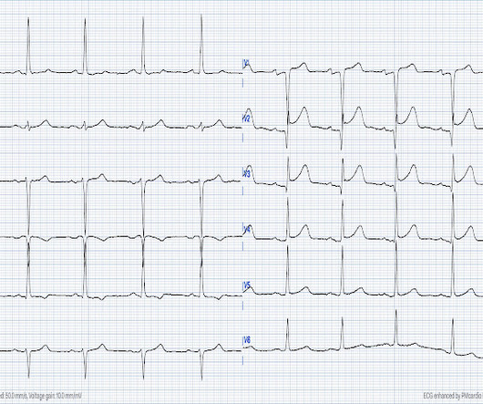
This EKG is diagnostic of transmural ischemia of the inferior wall. The Queen of Hearts disagrees, diagnosing OMI with high confidence: Case Continued: The EKG was not immediately recognized by the emergency provider, who ordered a CT scan to rule out aortic dissection at 1419. Lead I also shows reciprocal ST depression.
Objective The initial operation for type A aortic dissection has limitations, and there may be a need for reoperation in cases such as giant pseudoaneurysm formation and reduced blood supply to the distal vessels. In this study, we retrospectively analyzed the recorded data of 62 patients.
He interprets here: "This EKG is diagnostic of right bundle branch block and transmural ischemia of the anterior wall, most likely from an occlusion of the proximal LAD. The provider had sent the patient for an aortic dissection scan which had shown extremely heavy calcification of the LAD. It was recorded at 0530: What do you think?
To our knowledge, no studies have directly compared the right and left TRA for carotid artery stenting (CAS). The two groups exhibited similar patient characteristics, anatomical factors including aortic arch, and clinical outcomes. The mean age, percentage of male patients, and incidence of symptomatic cases were 73.9
FFR is obtained by dividing the pressure distal to the stenosis by the central aortic pressure, which is usually equal to the pressure proximal to the stenosis if there is no additional stenosis in between. indicates inducible ischemia while an FFR above 0.80 excludes ischemia in 90% of cases. Normal FFR is 1.0 in the study.
Post by Smith and Meyers Sam Ghali ( [link] ) just asked me (Smith): "Steve, do left main coronary artery *occlusions* (actual ones with transmural ischemia) have ST Depression or ST Elevation in aVR?" That said, complete LM occlusion would be expected to have subepicardial ischemia (STE) in these myocardial territories: STE vector 1.
There is ventricular hypertrophy in the absence of abnormal loading conditions, such as aortic stenosis, or hypertension, for example – of which the most common variant is Asymmetric Septal Hypertrophy. There is LBBB-like morphology with persistent patterns of subendocardial ischemia.
It was a 60yo with a history of stents to the circumflex and right coronary arteries, who presented with 9 hours of fluctuating central chest pain. 2] Here there is no posterior ST elevation, but the anterior ST depression is also less—so it is dynamic, confirming acute ischemia. But it is still STEMI negative.
A middle-aged male with h/o CAD and stents presented with typical chest pressure. An elderly patient with a ruptured abdominal aortic aneurysm: Formal ECG Interpretation (final read in the chart!) : "Inferior ST elevation, lead III, with reciprocal ST depression in aVL." This is a very common misread. What do you think?
On arrival his BP was 70s/40s, so an intra-aortic ballon pump was placed. This means that they occur shortly after onset of occlusion, but also may be the last remaining sign of ischemia after ST elevation resolves (after reperfusion). A repeat ECG was done: Obvious anterolateral wall STEMI. This is diagnostic of re-occlusion.
60-something with h/o MI and stents presented with chest pain radiating to the back and nausea/vomiting. There was concern for aortic dissection, so a CT was done and was negative. It was stented. The patient had a p rior h istory of MI + stents. More likely, these T waves probably reflect ischemia of uncertain age.
The first troponin returned at 0.099 ng/mL (elevated, consistent with Non-Occlusion MI) Providers were concerned with aortic dissection, so they order a chest aorta CT. This transmural ischemia, but not necessarily completed infarction (yet).
Ischemia often produces a straightening of the ST segment and sometimes upward convexity. At cath later the same day, a proximal 99% RCA culprit lesion was stented. And, even if there was acute aortic dissection — the dissection could result in occlusion of a coronary artery. Troponin T peaked at 4051 ng/L.
His initial high sensitivity troponin I returned at 1300 ng/L and given that his cardiac workup was otherwise unremarkable, a CT was obtained to evaluate for pulmonary embolism and aortic aneurysm or dissection but this too was unrevealing. Another EKG was also obtained. ECG at time 82 minutes: What do you think?
There was some question of whether the patient was having abdominal pathology, and she also had a history of aortic pathology, so a chest abd/pelvic with aorta angiogram was ordered. It was opened and stented. If this were ACUTE (vs. SUBACUTE) OMI, that would result in an undesirable delay.
We organize all of the trending information in your field so you don't have to. Join thousands of users and stay up to date on the latest articles your peers are reading.
You know about us, now we want to get to know you!
Let's personalize your content
Let's get even more personalized
We recognize your account from another site in our network, please click 'Send Email' below to continue with verifying your account and setting a password.


Let's personalize your content