This site uses cookies to improve your experience. To help us insure we adhere to various privacy regulations, please select your country/region of residence. If you do not select a country, we will assume you are from the United States. Select your Cookie Settings or view our Privacy Policy and Terms of Use.
Cookie Settings
Cookies and similar technologies are used on this website for proper function of the website, for tracking performance analytics and for marketing purposes. We and some of our third-party providers may use cookie data for various purposes. Please review the cookie settings below and choose your preference.
Used for the proper function of the website
Used for monitoring website traffic and interactions
Cookie Settings
Cookies and similar technologies are used on this website for proper function of the website, for tracking performance analytics and for marketing purposes. We and some of our third-party providers may use cookie data for various purposes. Please review the cookie settings below and choose your preference.
Strictly Necessary: Used for the proper function of the website
Performance/Analytics: Used for monitoring website traffic and interactions
6, 2025 Medtronic plc hasannounced it received CE ( Conformit Europenne ) Mark for the Harmony Transcatheter Pulmonary Valve (TPV) System, a minimally invasive alternative to open-heart surgery for congenital heartdisease patients with native or surgically repaired right ventricular outflow tract (RVOT).
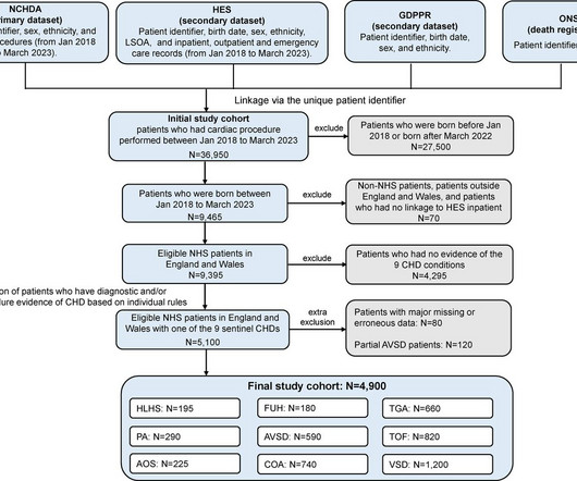
Background Infants with congenital heartdisease (CHD) are clinically vulnerable to cardiac deteriorations and intercurrent infections. We aimed to quantify the impact of health system disruptions during the COVID-19 pandemic, on their clinical outcomes and whether these differed by socioeconomic and ethnic subgroups.
Animal studies have shown that mice with TBX1 gene mutations have smaller left pulmonary arteries compared to wild type mice, defined by a reduced left pulmonary artery (LPA) to right pulmonary artery (RPA) ratio. A single study has shown this translates to humans with 22q11 and structurally normal hearts.
Right Heart Catheterization in Tetralogy of Fallot With the availability of high resolution echocardiographic images and Doppler echocardiography, role of cardiac catheterization has come down in tetralogy of Fallot and other congenital heartdiseases in general. Normal subjects have a value around 2.1.
Transcript of video: Hypoplastic Left Heart Syndrome is a very severe form of congenital heartdisease, in which, the left ventricle, aorta and mitral and aortic valves are hypoplastic and valves may be atretic as well. This is diagrammatic representation of hypoplastic left heart syndrome.
Purpose This study aims to evaluate deep learning (DL) denoising reconstructions for image quality improvement of Doppler ultrasound (DUS)-gated fetal cardiac MRI in congenital heartdisease (CHD). Diagnostic confidence was assessed for the atria, ventricles, foramen ovale, valves, great vessels, aortic arch, and pulmonary veins.
Expanding access to heartdisease detection is one of cardiology’s biggest challenges, and Finnish startup CardioSignal just raised $10M to address that challenge using one of the most accessible devices in the world – the smartphone.
This is the aortic valve in closed position and mitral valve also appears to be closed in position. You can also see the aortic override. So, when there is an aortic override, if the override of the aorta is less than 50%, you think of tetralogy of Fallot. Separation between the attachments of the aortic and mitral valve.
We describe a fetus with prenatal echocardiographic findings of BDA and right aortic arch mirror-image branching (RAA-MIB) combined with congenital heartdisease. According to the double arch theory, BDA is formed when the distal ends of the sixth pairs of primitive arches on the left and right sides have not regressed.
Like the heart chambers, there are four heart valves between each of the chambers. The pulmonary semilunar valve is between the right ventricle and the pulmonary trunk. The aortic semilunar valve is between the left ventricle and the aorta. Veins, on the other hand, return deoxygenated blood to the heart.
Look at the aortic outflow tract. The diagnostic coronary angiogram identified only minimal coronary artery disease, but there was a severely calcified, ‘immobile’ aortic valve. Aortic angiogram did not reveal aortic dissection. In fact, bedside ultrasound might even find severe aortic stenosis.
A good knowledge of the anatomy of the heart is needed for interpretation of images from each view. This becomes more difficult in complex congenital heartdiseases where the cardiac chamber positions and size may vary. This view images the heart from the base to apex long axis view.
Photo courtesy of Mount Sinai Health System milla1cf Thu, 02/22/2024 - 13:47 February 22, 2024 — Ismail El-Hamamsy , MD, PhD, Director of Aortic Surgery for the Mount Sinai Health System and the Mount Sinai Randall B. The aortic valve controls blood flow from the heart into the aorta, the main artery that feeds blood to most of the body.
Objectives This study aimed to evaluate the prognostic value of coronary microvascular dysfunction (CMD) at long term after transcatheter aortic valve implantation (TAVI) and to explore its relationship with extravalvular cardiac damage (EVCD). CMD was defined as IMR angio ≥30 units. CMD was defined as IMR angio ≥30 units.
Transcript of video: Tetralogy of Fallot is one of the commonest cyanotic congenital heartdiseases. One is ventricular septal defect, second is overriding aorta, third is pulmonary stenosis, usually right ventricular outflow tract stenosis and associated right ventricular hypertrophy. Right to left shunt is also visible.
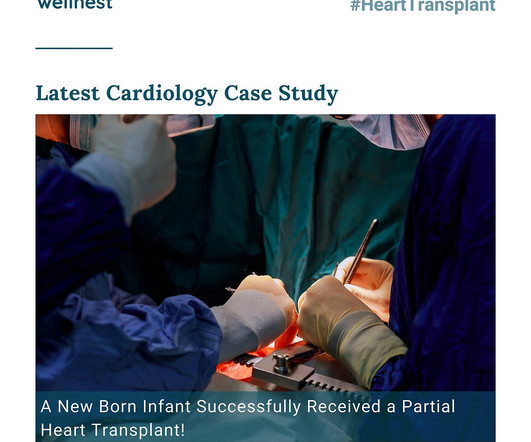
Unlike traditional approaches relying on non-viable cadaver grafts, this procedure involved the transplantation of a portion of the heart, specifically containing the aorta and pulmonary valves, sourced from an infant donor upon cardiac death. Finally, the pulmonary root was transplanted, completing the intricate procedure.
While the first one may radiate to the axilla and base, but usually not into the neck, it does reflect both aortic outflow obstruction and mitral regurgitation in patients with a large gradient. On the other hand, the murmur in valvular aortic stenosis does not change substantially or decreases slightly following the Valsalva maneuver.
Program Designations Access and Publications (A&P) 1 Participant User File (PUF) 2 Task Force on Funded Research (TFR) 3 Special Projects 4 Adult Cardiac Surgery Database Lead Author Title Publication Date William Keeling 2 National Trends in Emergency Coronary Artery Bypass Grafting European Journal of Cardiothoracic Surgery October 2023 Jake (..)
In pregnant women with significant heartdisease : A quick LSCS or a potentially prolonged natural delivery,which is more safe ? For women with significant heartdisease (e.g., In women with significant heartdisease, the physiological demands of labor (e.g., Is LSCS really more safe ?
Larger shunt volume means less blood exiting the left ventricle through the aortic valve and lower cardiac output. Rupture can be either free wall rupture (causing tamponade) or septal rupture, causing ventricular septal defect with left to right flow and resulting pulmonary edema and shock. When did the MI begin? When was the VSR?
We organize all of the trending information in your field so you don't have to. Join thousands of users and stay up to date on the latest articles your peers are reading.
You know about us, now we want to get to know you!
Let's personalize your content
Let's get even more personalized
We recognize your account from another site in our network, please click 'Send Email' below to continue with verifying your account and setting a password.


Let's personalize your content