This site uses cookies to improve your experience. To help us insure we adhere to various privacy regulations, please select your country/region of residence. If you do not select a country, we will assume you are from the United States. Select your Cookie Settings or view our Privacy Policy and Terms of Use.
Cookie Settings
Cookies and similar technologies are used on this website for proper function of the website, for tracking performance analytics and for marketing purposes. We and some of our third-party providers may use cookie data for various purposes. Please review the cookie settings below and choose your preference.
Used for the proper function of the website
Used for monitoring website traffic and interactions
Cookie Settings
Cookies and similar technologies are used on this website for proper function of the website, for tracking performance analytics and for marketing purposes. We and some of our third-party providers may use cookie data for various purposes. Please review the cookie settings below and choose your preference.
Strictly Necessary: Used for the proper function of the website
Performance/Analytics: Used for monitoring website traffic and interactions
Transcript of the video: Closure line of aortic valve on M-Mode echocardiogram, is seen as central line, while in bicuspid aortic valve, it is an eccentric closure, nearer to one of the walls of the aorta. This eccentricity of closure of the aortic valve leaflets, can be calculated using what is known as eccentricity index.
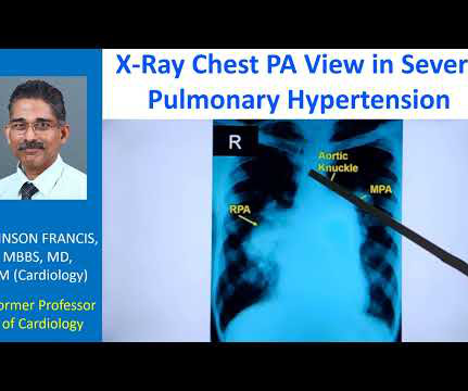
The striking finding is the huge enlargement of the right pulmonary artery, almost aneurysmal dilatation of right pulmonary artery. Main pulmonary artery is also grossly dilated. And you can see left pulmonary artery shadow and rest of it is not seen here. This is the aortic knuckle. This could be an end on view.
Transcript of video: Hypoplastic Left Heart Syndrome is a very severe form of congenital heart disease, in which, the left ventricle, aorta and mitral and aortic valves are hypoplastic and valves may be atretic as well. A Gore-Tex tube is used and this maintains, this is a Blalock-Taussig shunt, which maintains pulmonary circulation.
This is the schematic diagram of the heart in which you can see right atrium, right ventricle, left atrium, left ventricle, aorta and pulmonary artery. Unlike the valves on the left side like the mitral and aortic, right sided valves can have some leak. Similarly, another right sided valve is the pulmonary valve.
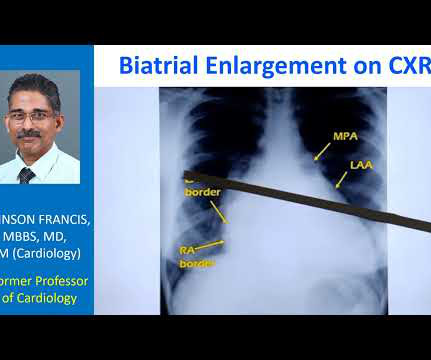
Normally, the main pulmonary artery segment will be concave and left atrial appendage region also will be not prominent. So that is why we see straightening of left border, typically heard of in mitral stenosis with left atrial enlargement and mild pulmonary hypertension. Those are not very clear in this picture.
This is the aortic valve in closed position and mitral valve also appears to be closed in position. You can also see the aortic override. So, when there is an aortic override, if the override of the aorta is less than 50%, you think of tetralogy of Fallot. Separation between the attachments of the aortic and mitral valve.
Transcript of the video: Eisenmenger syndrome is an important complication of large left to right shunts which develop later due to development of pulmonary vascular obstructive disease and severe pulmonary hypertension. So in ASD Eisemenger, suprasystemic pulmonary hypertension is possible. But, leave that alone.
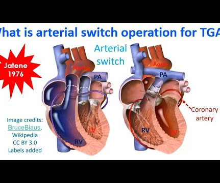
That is, right ventricle is connecting to aorta, and left ventricle to pulmonary artery. That is, pulmonary artery is transposed over to the right ventricle, and aorta over to the left ventricle, so that normal anatomy is restored. In dextro transposition of great arteries or D-TGA, there is ventriculoarterial discordance.
Leaving a fenestration in the interatrial septum during a Fontan repair is useful in relieving the central venous congestion when pulmonary blood flow is driven by venous pressure in Fontan repair. They noted that benefit was more pronounced if mean pulmonary arterial pressure was 13 mm Hg or more, as expected.
So it will not produce a true LV to aorta pullback tracing, which is required in cases like aortic stenosis. While standard pigtail catheter is mainly used on the left side, Grollman PA is a catheter used for pulmonary angiography, on the right side. When the tip is in the left ventricle, this region will be in the aorta sometimes.
Usual structures imaged in this view are the right ventricular free wall and outflow region, interventricular septum, aorta, and aortic valve, left ventricular outflow tract, anterior and posterior mitral leaflets, left ventricular cavity, posterior wall of left ventricle and left atrium. Colour flow shows the flow in pulmonary artery.
One is ventricular septal defect, second is overriding aorta, third is pulmonary stenosis, usually right ventricular outflow tract stenosis and associated right ventricular hypertrophy. You can see the ventricular septal defect and aortic over ride. Pulmonary stenosis, which is usually right ventricular outflow tract stenosis.
I has to be done quite early in life before the left ventricular muscle mass regressed due to the lower load of the pulmonary circulation. Rare long term problems include narrowing of the pulmonary artery and aortic regurgitation due to aortic root enlargement.
We organize all of the trending information in your field so you don't have to. Join thousands of users and stay up to date on the latest articles your peers are reading.
You know about us, now we want to get to know you!
Let's personalize your content
Let's get even more personalized
We recognize your account from another site in our network, please click 'Send Email' below to continue with verifying your account and setting a password.


Let's personalize your content