This site uses cookies to improve your experience. To help us insure we adhere to various privacy regulations, please select your country/region of residence. If you do not select a country, we will assume you are from the United States. Select your Cookie Settings or view our Privacy Policy and Terms of Use.
Cookie Settings
Cookies and similar technologies are used on this website for proper function of the website, for tracking performance analytics and for marketing purposes. We and some of our third-party providers may use cookie data for various purposes. Please review the cookie settings below and choose your preference.
Used for the proper function of the website
Used for monitoring website traffic and interactions
Cookie Settings
Cookies and similar technologies are used on this website for proper function of the website, for tracking performance analytics and for marketing purposes. We and some of our third-party providers may use cookie data for various purposes. Please review the cookie settings below and choose your preference.
Strictly Necessary: Used for the proper function of the website
Performance/Analytics: Used for monitoring website traffic and interactions
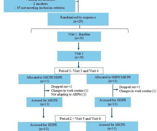
Objective In this randomised two-period crossover trial, the objective was to compare acute changes in arterial distensibility between high-intensity interval physical exercise (HIIPE) and moderate-intensity continuous physical exercise (MICPE) sessions in subjects with elevated blood pressure (BP).
Valvular heart disease, including calcific or degenerative aortic stenosis (AS), is increasingly prevalent among the older adult population. Over the last few decades, treatment of severe AS has been revolutionised following the development of transcatheter aortic valve replacement (TAVR).
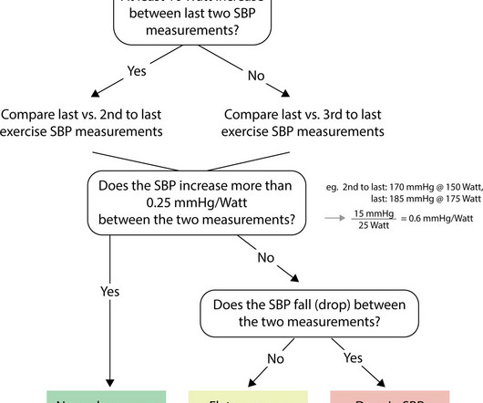
Aims Exercise testing remains underused in patients with aortic stenosis (AS), partly due to concerns about an exercise-induced drop in systolic blood pressure (SBP). We aimed to study the SBP response to exercise in patients with severe symptomatic AS prior to surgery and 1 year postoperatively.
Aortic coarctation (AoC) is a common congenital heart defect, affecting 5%8% of patients with structural congenital anomalies. In these patients, hypertension is associated to renin-angiotensin system activation, residual aortic arch abnormalities, and impaired aortic elasticity.
BackgroundThe potential impact of exercise on valvular function and aortic diameters in patients with a bicuspid aortic valve remains unclear. Echocardiography was used to assess aortic stenosis or aortic regurgitation and to measure diameters at the sinuses of Valsalva and ascending aorta. and 20.0%, respectively.
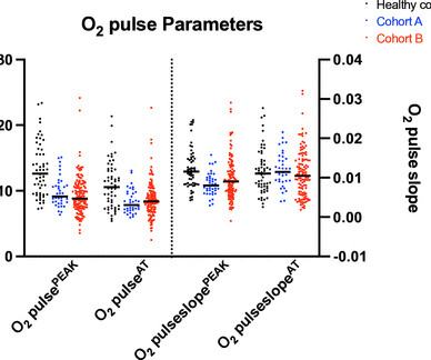
We propose that the slope of the O 2 pulse curve may be more reflective of SV during exercise. Results In cohort A, peak aortic flow was moderately and significantly associated with O 2 pulseslope PEAK (r=0.47, p=0.02). However, neither absolute O 2 pulse AT nor O 2 pulse PEAK was significantly associated with peak aortic flow.
Transcatheter aortic valve replacement (TAVR) is a relatively new treatment method for aortic stenosis (AS) and has been demonstrated to be suitable for patients with varying risk levels.
Objective Decreased proximal aortic distensibility (AD) is known to significantly predict all-cause mortality and cardiovascular events among individuals without overt cardiovascular disease. Ascending aortic diameters were measured simultaneously with pulse pressure. Aortic strain, AD and aortic stiffness index were calculated.
Lp(a) is emerging as an important, yet under-recognized, potential risk factor for cardiovascular disease due to its ability to promote the development of plaques within artery walls, clot formation and aortic valve calcification. 2022 Aug, 80 (9) 934946 Kronenberg F.
Optimal administration of exercise or intravenous drugs may reveal hemodynamic abnormalities under stress without posing an invasive risk. Therefore, the implementation of stress echocardiography is recommended for determining interventional indications and risk stratification in mitral regurgitation and aortic stenosis.
If you’ve been diagnosed with aortic stenosis, you might have come across the term TAVR. Understanding Aortic Stenosis The aortic valve regulates blood flow from your heart’s main pumping chamber to the rest of your body. In aortic stenosis, the valve leaflets stiffen and narrow, restricting blood flow.
Publication date: Available online 19 August 2024 Source: The American Journal of Cardiology Author(s): Chidiogo Orizu, Mawra Jha, Lana Myerson, Zhiyong J. Dong, Ulf Neisius, Inbar McCarthy, Dharshan Lakshminarayan, Warren J. Manning, Connie W.
24: Joint American College of Cardiology/Journal of the American College of Cardiology Late-Breaking Clinical Trials (Session 402) Saturday, April 6 9:30 – 10:30 a.m.
The patient was referred for an exercise nuclear study and did 11 min on the Bruce protocol without angina or ischaemic ECG changes. Aortic dissection Sinus of Valsalva aneurysm Anomalous coronary artery Unroofed. He did not smoke or use alcohol or illicit drugs. There was no pertinent family history.
Background:Heart failure with preserved left ventricular ejection fraction (LVEF) prevails in subjects who complained of exercise intolerance, which is characterized by elevated left ventricular filling pressure (LVFP). Correlation matrix analysis showed aortic AI, Tau, and RRA AI and all three vessels vFFR correlated with LVEDP.
MC1R recessive yellow mice showed blunted hypertrophic response to transverse aortic constrictioninduced pressure overload and exercise training. Mice were phenotyped for cardiac structure and function by echocardiography, histology, and quantitative PCR analysis.
But, still for an academic exercise, we will try. This is the aortic valve in closed position and mitral valve also appears to be closed in position. You can also see the aortic override. So, when there is an aortic override, if the override of the aorta is less than 50%, you think of tetralogy of Fallot.
The typical pain of cardiac origin is a central chest pain which occurs on walking or other forms of exercise, known as effort angina. Pain of aortic dissection is tearing type and is most severe in the beginning. Still some general observations are possible regarding chest pain originating from the heart.
We are blessed with 4 heart valves – 2 on the left side which are known as the mitral and aortic valves and 2 on the right side – the tricuspid and pulmonary valves. He is intolerant of exercise for the same reason. He swells up with swelling of the legs and the abdomen because of all this volume overload.
Furthermore, she denies any hydration since conclusion of exercise. There is ventricular hypertrophy in the absence of abnormal loading conditions, such as aortic stenosis, or hypertension, for example – of which the most common variant is Asymmetric Septal Hypertrophy.
1, 2024 — Researchers at UTHealth Houston have identified genetic variants linked to a rare form of bicuspid aortic valve disease that affects young adults and can lead to dangerous and potentially life-threatening aortic complications. tim.hodson Wed, 09/04/2024 - 15:53 Sept.
BackgroundThe prognostic value of serial exercise echocardiography (EEC) in asymptomatic severe aortic stenosis is unknown. Journal of the American Heart Association, Ahead of Print.
Edwards has entered into an agreement to acquire JenaValve Technology , a pioneer in the transcatheter treatment of aortic regurgitation (AR), a deadly disease that impacts a significant and growing population and is largely untreated today. JenaValve presented positive results of its U.S.
Venn diagram highlighting the main similarities and differences between heart failure with preserved ejection fraction (HFpEF) and aortic stenosis with preserved ejection fraction (ASpEF). This study aimed to provide a non-invasive, comparative analysis of ASpEF versus HFpEF at rest and during exercise. vs. controls).
Written by Pendell Meyers A woman in her 20s with connective tissue disorder and history of aortic root and valve repair presented with palpitations. Further history revealed she had new onset atrial flutter soon after her aortic surgery, and was put on flecainide approximately 1 month ago. Here is her triage ECG: What do you think?
You can see the ventricular septal defect and aortic over ride. Hemodynamic effects of pulmonary regurgitation include chronic right ventricular volume overload, right ventricular dysfunction and exercise intolerance. And, usually this done, Blalock-Taussig shunt, classic shunt, opposite to the side of the aortic arch.
He visited an outpatient clinic for it and an echocardiogram and exercise stress test was normal. As his pain was very severe, emergency physicians concerned of aortic dissection and ordered a thoracic CT scan. He has 40 packs-year of smoking history. There was no premature cardiovascular diseases or sudden death in his family.
Sent by anonymous, written by Pendell Meyers A male in his teens presented with complaints of chest discomfort and dyspnea beginning while exercising but without obvious injury. He immediately stopped exercising and symptoms started to improve. CT angiogram chest: no aortic dissection or pulmonary embolism.
BACKGROUND:Recent guidelines redefined exercise pulmonary hypertension as a mean pulmonary artery pressure/cardiac output (mPAP/CO) slope >3 mm Hg·L−1·min−1. cm2underwent cardiopulmonary exercise testing with echocardiography. Peak aortic velocity (odds ratio [OR] per SD, 1.48;P=0.036),
Objective To characterise cardiac remodelling, exercise capacity and fibroinflammatory biomarkers in patients with aortic stenosis (AS) with and without diabetes, and assess the impact of diabetes on outcomes. to 4.00; p=0.037).
Background Knowledge about how patients with symptomatic aortic stenosis (AS) perform on cardiopulmonary exercise testing (CPET) is sparse. Since exercise testing in patients with symptomatic AS is not advised, submaximal parameters could be of special interest.
13 Coronary artery calcification (CAC) and aortic stiffness are also associated with self-reported sleep duration. 4 Many environmental and lifestyle factors can throw our circadian system out of whack, which is why being prudent about sleep, diet, and exercise are crucial.
Below are some of the most common causes of heart murmurs: Increased Blood Flow: Innocent heart murmurs often occur when there is an increase in blood flow, such as during pregnancy, exercise, fever, or growth spurts in children. The aortic valve and mitral valve are two of the most common valves affected by heart murmurs.
If LV contraction interferes with coronary blood flow, a patient with severe LV dysfunction gains some advantage as systolic blood flow can happen more easily, and myocardium is perfused better, provided his aortic systolic pressure not too low enough. How common is angina in DCM ? Angina in DCM is an exception despite elevated LVEDP.
We organize all of the trending information in your field so you don't have to. Join thousands of users and stay up to date on the latest articles your peers are reading.
You know about us, now we want to get to know you!
Let's personalize your content
Let's get even more personalized
We recognize your account from another site in our network, please click 'Send Email' below to continue with verifying your account and setting a password.


Let's personalize your content