This site uses cookies to improve your experience. To help us insure we adhere to various privacy regulations, please select your country/region of residence. If you do not select a country, we will assume you are from the United States. Select your Cookie Settings or view our Privacy Policy and Terms of Use.
Cookie Settings
Cookies and similar technologies are used on this website for proper function of the website, for tracking performance analytics and for marketing purposes. We and some of our third-party providers may use cookie data for various purposes. Please review the cookie settings below and choose your preference.
Used for the proper function of the website
Used for monitoring website traffic and interactions
Cookie Settings
Cookies and similar technologies are used on this website for proper function of the website, for tracking performance analytics and for marketing purposes. We and some of our third-party providers may use cookie data for various purposes. Please review the cookie settings below and choose your preference.
Strictly Necessary: Used for the proper function of the website
Performance/Analytics: Used for monitoring website traffic and interactions
BackgroundAbdominal aortic calcification (AAC), an early indicator of abdominal aortic wall atherosclerosis, is a marker of subclinical atherosclerosis and a predictive factor for vascular-associated morbidity and mortality. These outcomes are driven by inflammatory processes.
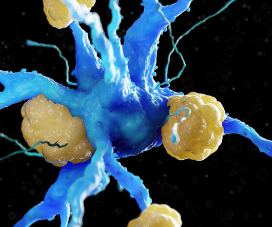
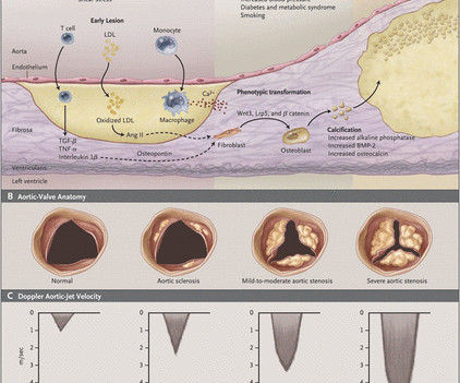
Getty Images milla1cf Fri, 06/07/2024 - 15:10 June 7, 2024 — Calcific aortic valve disease (CAVD) is the major heart valve disease that afflicts nearly 10 million patients globally with an annual mortality exceeding 100,000, and the numbers continue to rise. These findings appear online in the journal Trends in Molecular Medicine.
Aortic stenosis (AS) due to fibrosis and calcification of the aortic valve is a hazardous component of cardiovascular disease burden—after developing symptomatic AS, patients survive for an average of less than 2 years without treatment.
Objective Aortic stenosis (AS) shares pathophysiological similarities with atherosclerosis including active inflammation. CT attenuation of perivascular adipose tissue provides a measure of vascular inflammation that is linked to prognosis and has the potential to be applied to the aortic valve. HU, p=0.099).
Introduction The presence of non-coronary atherosclerosis (NCA) in patients with coronary artery disease is associated with a poor prognosis. We have studied whether NCA is also a predictor of poorer outcomes in patients undergoing coronary artery bypass grafting (CABG).
Hutchinson-Gilford Progeria Syndrome (HGPS) is an ultra-rare genetic premature aging disease that is historically fatal in teenage years, secondary to severe accelerated atherosclerosis. With this longer lifespan, calcific aortic stenosis (AS) was identified as an emerging critical risk factor for cardiac death in older patients.
Recent studies implicate EndMT in atherosclerosis development. Diabetes is associated with both EndMT as well as accelerated atherosclerosis. Experimental evidence supports the use of epigenetic drugs that act on relevant epigenetic mechanisms to attenuate atherosclerosis.
BackgroundUltraviolet B (UV‐B) irradiation is an effective treatment for human cutaneous disorders and was shown to reduce experimental atherosclerosis by attenuating immunoinflammatory responses. Our findings indicate that a novel 282 nm UV‐B phototherapy could be an attractive approach to treat atherosclerosis.
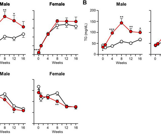
Atherosclerosis was evaluated at the endpoint of experiments. fold increase) apoE KO rabbits exhibited a significantly augmented aortic lesion area compared to WT controls. cholesterol for 16 weeks. Plasma lipid levels, lipoproteins, and apolipoproteins were analyzed. Notably, both male (2.1-fold fold increase) and female (1.6-fold
Aortic diseases such as atherosclerosis, aortic aneurysms, and aortic stiffening are significant complications that can have significant impact on end-stage cardiovascular disease. Hypertension, Ahead of Print.
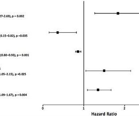
This study examined whether HGF is associated with ECC in the aortic valve (AVC), mitral annulus (MAC), ascending thoracic aorta and descending thoracic aortic (DTAC). However, knowledge is limited on the role HGF may play in extracoronary calcification (ECC).
Our perspectives on aortic stenosis (AS) are changing. The pathophysiology of calcific AS (CAS) is complex, yet can be characterised similarly to that of atherosclerosis. In addition, their integration with cardiovascular MRI can provide accurate risk stratification, aiding aortic valve replacement decision making.
Introduction The presence of abdominal aortic calcification (AAC) is strongly linked to the development of atherosclerosis and the incidence of morbidity and mortality related to cardiovascular diseases (CVD). Urinary albumin creatinine ratio (UACR) was found related with the increased risk of CVD.
BackgroundChronic inflammatory disease (CID) accelerates atherosclerosis and the development of aortic stenosis. Data on long‐term outcomes after transcatheter aortic valve implantation (TAVI) in those patients are missing.
Lp(a) is emerging as an important, yet under-recognized, potential risk factor for cardiovascular disease due to its ability to promote the development of plaques within artery walls, clot formation and aortic valve calcification. 2022 Aug, 80 (9) 934946 Kronenberg F.
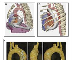
Transcatheter aortic valve replacement (TAVR) is increasing in popularity for symptomatic severe aortic stenosis. Transfemoral arterial route is the most commonly used approach for TAVR, also known as TAVI or transcatheter aortic valve implantation.
years [SD, 4.1]) from the ARIC (Atherosclerosis Risk in Communities) study, we measured segment‐specific PWVs: heart–carotid, heart–femoral, carotid–femoral, heart–ankle, brachial–ankle, and femoral–ankle.
BackgroundAortic stenosis has pathophysiological similarities with atherosclerosis, including the deposition of cholesterol‐containing lipoproteins. The resulting cholesterol crystals activate the NLRP3 (NOD‐like receptor protein 3) inflammasome, leading to inflammation and cardiovascular diseases.
Background and Objectives:Left subclavian artery (LSA) is more prone to atherosclerosis than the right one. The study was designed to investigate whether aortic arch types (AAT) was associated with the lateralization of subclavian artery stenosis (SAS).Methods:In Stroke, Volume 56, Issue Suppl_1 , Page ATP293-ATP293, February 1, 2025.
Objective Cell division cycle 42 (CDC42) regulates CD4 + T -cell differentiation and participates in vascular stiffness and atherosclerosis and is involved in the progression of Stanford type B aortic dissection (TBAD).
The Queen of Hearts disagrees, diagnosing OMI with high confidence: Case Continued: The EKG was not immediately recognized by the emergency provider, who ordered a CT scan to rule out aortic dissection at 1419. There is an area of dense white in the middle of the circle consistent with atherosclerosis. The blue circle shows the LCx.
BACKGROUND:Abdominal aortic aneurysm (AAA) is a potentially life-threatening vascular condition, but approved medical therapies to prevent AAA progression and rupture are currently lacking. GM3 content and ST3GAL5 expression were decreased in abdominal aortic vascular SMCs in patients with AAA and an AAA mouse model.
1,2 ASCVD causes or contributes to conditions that include coronary artery disease (CAD), cerebrovascular disease, and peripheral vascular disease (inclusive of aortic aneurysm).3 1,6 Until recently atherosclerosis has been thought of as the result of passive lipid accumulation in the vessel wall. 4 In the U.S.
This study compared the prognostic value of quantified thoracic artery calcium (TAC) including aortic arch on chest CT and coronary artery calcium (CAC) score on ECG-gated cardiac CT. TAC is defined as calcification in the ascending aorta, aortic arch and descending aorta on chest CT.
BACKGROUND:High circulating levels of Lp(a) (lipoprotein[a]) increase the risk of atherosclerosis and calcific aortic valve disease, affecting millions of patients worldwide.
Genome-wide association and Mendelian randomisation studies have identified lipoprotein(a) (Lp[a]) as an emerging risk factor for calcific aortic stenosis and a causal risk factor for atherosclerotic cardiovascular disease (ASCVD) in different ethnicities. HEART UK recommends Lp(a) measurement in specific ‘at-risk’ cohorts.
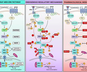
We then summarize current knowledge of the roles played by ERK in the development and progression of cardiac and vascular disorders, including atherosclerosis, myocardial infarction, cardiac hypertrophy, heart failure, and aortic aneurysm.
The fundamental characteristic of atherosclerosis is when a cholesterol particle becomes trapped in the artery wall. It is the inflammatory response to this particle retention that causes the formation of atherosclerosis 1. Lipoprotein particles entering the subintimal space causing atherosclerosis. CE = Cholesterol Ester.
The EGFR (epidermal growth factor receptor) has been shown to mediate inflammatory vascular diseases, including atherosclerosis and aortic aneurysm. BACKGROUND:Multiple pathways and factors are involved in the rupture of intracranial aneurysms.
A 69‐year‐old woman with a history of lung cancer, hypertension, chronic tobacco use, atherosclerosis, and known calcified plaque at the left carotid bifurcation on dual antiplatelet therapy presented with acute onset of expressive aphasia and right hemiparesis due to acute left CCAO.
Lipoprotein(a) (Lp[a]) can improve the accuracy of assessment of atherosclerotic cardiovascular disease and the risk of aortic valve stenosis. Currently, there is no specific treatment to lower its circulating concentration. Raised Lp(a) is a feature of familial hypercholesterolaemia.
Repeat CTA head and neck demonstrated multifocal intracranial atherosclerosis with marked stenosis of the left V4 segment. However, marked stenosis at V2 appeared to be owed to external compression from facet arthrosis rather than atherosclerosis. He also had moderate stenosis of the right V4 segment.
Cardiology noted there was no STEMI criteria and the first troponin was in the normal range (25ng/L, with normal <26), so alternate diagnoses were considered and the patient was sent for CT to rule out aortic dissection. 4] CT revealed no dissection but extensive coronary atherosclerosis. In a study last year, 14.4%
Mechanistically, HEG1 knockdown prevented s-flow–induced KLF2/4 (Krüppel-like factor 2/4) expression by regulating its intracellular binding partner KRIT1 (Krev interaction trapped protein 1) and the MEKK3-MEK5-ERK5-MEF2 pathway in human aortic endothelial cells. Circulation, Ahead of Print.
The amount of calcium in the blood vessels (known as arterial calcification), a marker of subclinical atherosclerosis, is higher in people with a short sleep duration. 13 Coronary artery calcification (CAC) and aortic stiffness are also associated with self-reported sleep duration.
Attendees, including hundreds of health professionals, gained access to the latest knowledge and developments in the field, from exclusive insights from one of the foremost authorities on atherosclerosis, Dr. Peter Libby, to innovations like new therapeutic agents and exciting advancements in renal protection.
We organize all of the trending information in your field so you don't have to. Join thousands of users and stay up to date on the latest articles your peers are reading.
You know about us, now we want to get to know you!
Let's personalize your content
Let's get even more personalized
We recognize your account from another site in our network, please click 'Send Email' below to continue with verifying your account and setting a password.


Let's personalize your content