This site uses cookies to improve your experience. To help us insure we adhere to various privacy regulations, please select your country/region of residence. If you do not select a country, we will assume you are from the United States. Select your Cookie Settings or view our Privacy Policy and Terms of Use.
Cookie Settings
Cookies and similar technologies are used on this website for proper function of the website, for tracking performance analytics and for marketing purposes. We and some of our third-party providers may use cookie data for various purposes. Please review the cookie settings below and choose your preference.
Used for the proper function of the website
Used for monitoring website traffic and interactions
Cookie Settings
Cookies and similar technologies are used on this website for proper function of the website, for tracking performance analytics and for marketing purposes. We and some of our third-party providers may use cookie data for various purposes. Please review the cookie settings below and choose your preference.
Strictly Necessary: Used for the proper function of the website
Performance/Analytics: Used for monitoring website traffic and interactions
Presentation title: QUALITY OF LIFE ASSESSMENT AT 30-DAYS FOLLOW-UP OF THE VALVOSOFT PIVOTAL STUDY ON SEVERE AORTICVALVE STENOSE PATIENTS" (control number 16930) Time and date: April 8, 2024 - 9:32 a.m.–9:42 A summary of the poster session will be published on the Journal of the American College of Cardiology’s website.
Food and Drug Administration (FDA) has granted 510(k) clearance for its first-of-a-kind, AI-powered AISAP CARDIO point-of-care ultrasound (POCUS) software platform. We know that structural heart disease and heart failure are the leading causes of hospitalization and morbidity in the U.S.
24: Joint American College of Cardiology/Journal of the American College of Cardiology Late-Breaking Clinical Trials (Session 402) Saturday, April 6 9:30 – 10:30 a.m.
Smith comment: This patient did not have a bedside ultrasound. Had one been done, it would have shown a feature that is apparent on this ultrasound (however, this patient's LV function would not be as good as in this clip): This is recorded with the LV on the right. Look at the aortic outflow tract. What should be done?
During echocardiography, a transducer transmits the ultrasound beam towards the heart. Planimetry of mitral valve area can be obtained in parasternal short axis view in case of mitral stenosis. It is used in the emergency department, at bedside, in the intensive care unit as well as in the operating room.
They found non-obstructive CAD, with only a 20% stenosis of OM2 and 10% RCA. The next morning the patient went for his routine echocardiogram, where the operator noticed a dilated aortic root at 5.47 cm with severe aortic insufficiency. Beware a negative Bedside ultrasound. No acute culprit. He was admitted to cardiology.
Venn diagram highlighting the main similarities and differences between heart failure with preserved ejection fraction (HFpEF) and aorticstenosis with preserved ejection fraction (ASpEF). Patients with ASpEF eligible for transcatheter aorticvalve replacement ( n = 125) also performed cardiac computed tomography (CT).
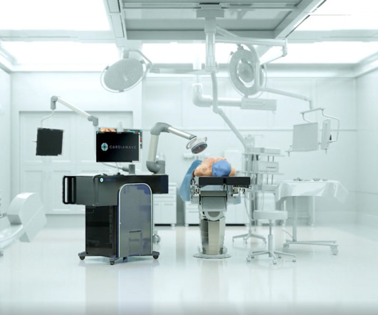
28, 2024 — Cardiawave SA, developer of Valvosoft Non-Invasive Ultrasound Therapy (NIUT) device for treating severe symptomatic calcific aorticstenosis (CAS), announced that the device met the primary endpoint in its pivotal study and improved or stabilized heart failure symptoms for 80.5 tim.hodson Thu, 11/07/2024 - 10:19 Oct.
I suspect pulmonary edema, but we are not given information on presence of B-lines on bedside ultrasound, or CXR findings. Smith : "decompensation" of aorticstenosis might have initiated this entire cascade. What "initiates" the aorticstenosis cascade? We certainly know that there is hypoxia.
We organize all of the trending information in your field so you don't have to. Join thousands of users and stay up to date on the latest articles your peers are reading.
You know about us, now we want to get to know you!
Let's personalize your content
Let's get even more personalized
We recognize your account from another site in our network, please click 'Send Email' below to continue with verifying your account and setting a password.


Let's personalize your content